• 文献检索
  • 文档翻译
  • 深度研究
  • 学术资讯
  • Suppr Zotero 插件Zotero 插件
  • 邀请有礼
  • 套餐&价格
  • 历史记录
应用&插件
Suppr Zotero 插件Zotero 插件浏览器插件Mac 客户端Windows 客户端微信小程序
定价
高级版会员购买积分包购买API积分包
服务
文献检索文档翻译深度研究API 文档MCP 服务
关于我们
关于 Suppr公司介绍联系我们用户协议隐私条款
关注我们

Suppr 超能文献

核心技术专利:CN118964589B侵权必究
粤ICP备2023148730 号-1Suppr @ 2026

文献检索

告别复杂PubMed语法,用中文像聊天一样搜索,搜遍4000万医学文献。AI智能推荐,让科研检索更轻松。

立即免费搜索

文件翻译

保留排版,准确专业,支持PDF/Word/PPT等文件格式,支持 12+语言互译。

免费翻译文档

深度研究

AI帮你快速写综述,25分钟生成高质量综述,智能提取关键信息,辅助科研写作。

立即免费体验

光学相干断层扫描:洞察冠状动脉的眼睛。

Optical Coherence Tomography: An Eye Into the Coronary Artery.

作者信息

Gupta Ankush, Shrivastava Abhinav, Vijayvergiya Rajesh, Chhikara Sanya, Datta Rajat, Aziz Atiya, Singh Meena Daulat, Nath Ranjit Kumar, Kumar J Ratheesh

机构信息

Department of Cardiology, Military Hospital Jaipur, Jaipur, India.

Department of Cardiology, Dr Ram Manohar Lohia (RML) Hospital & Atal Bihari Vajpayee Institute of Medical Sciences (ABVIMS), New Delhi, India.

出版信息

Front Cardiovasc Med. 2022 May 11;9:854554. doi: 10.3389/fcvm.2022.854554. eCollection 2022.

DOI:10.3389/fcvm.2022.854554
PMID:35647059
原文链接:https://pmc.ncbi.nlm.nih.gov/articles/PMC9130606/
Abstract

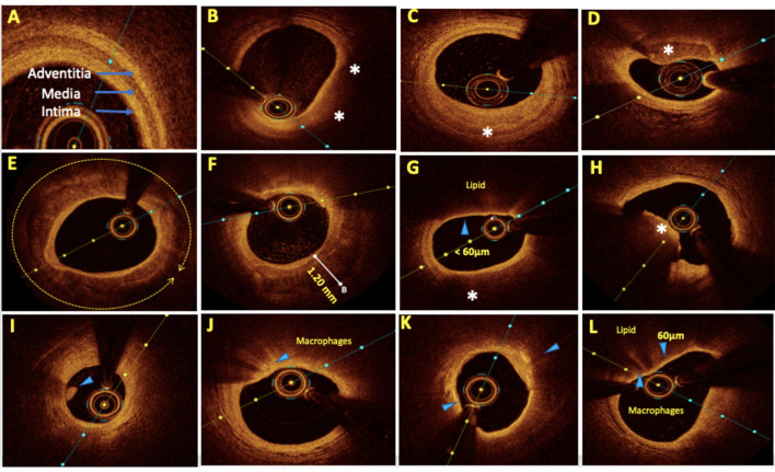
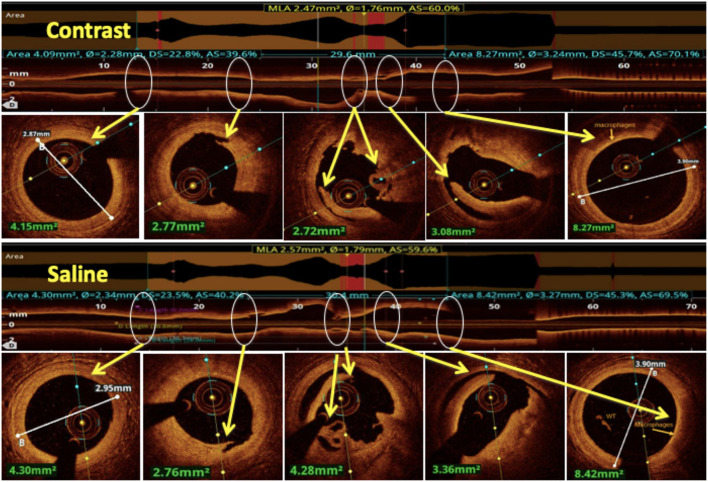
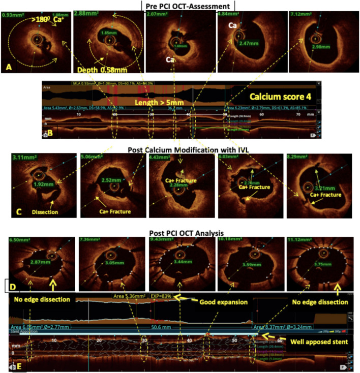
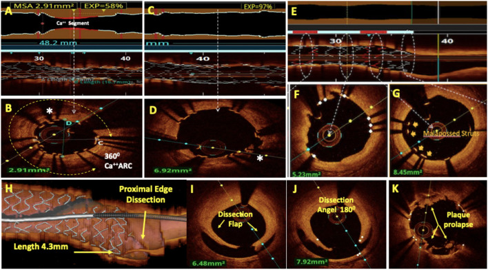
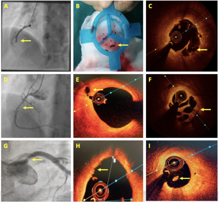
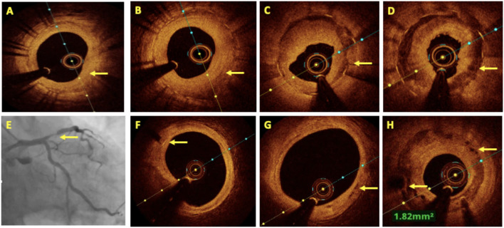
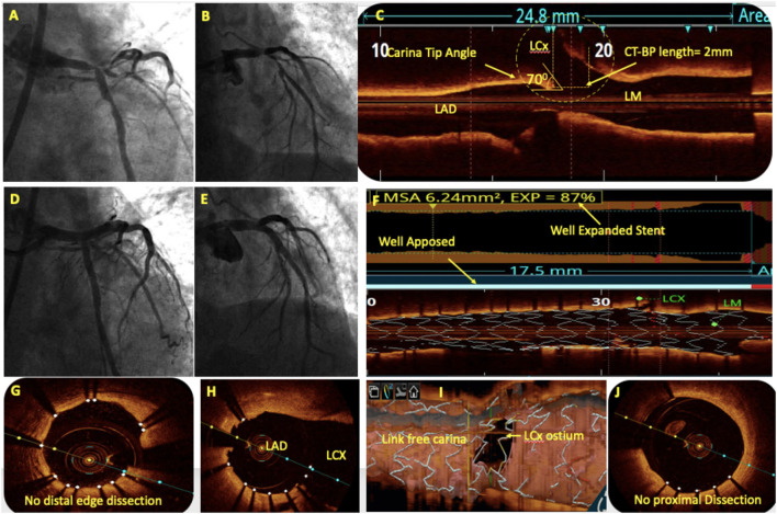
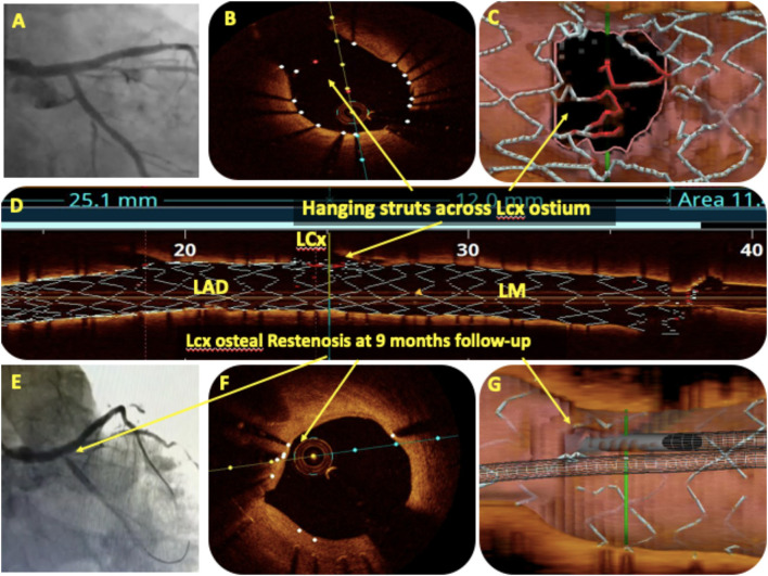
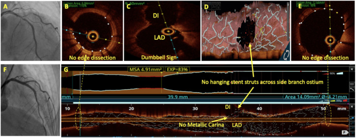
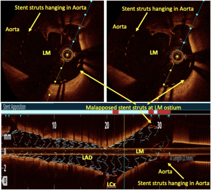
Optical coherence tomography (OCT) is slowly but surely gaining a foothold in the hands of interventional cardiologists. Intraluminal and transmural contents of the coronary arteries are no longer elusive to the cardiologist's probing eye. Although the graduation of an interventionalist in imaging techniques right from naked eye angiographies to ultrasound-based coronary sonographies to the modern light-based OCT has been slow, with the increasing regularity of complex coronary cases in practice, such a transition is inevitable. Although intravascular ultrasound (IVUS) due to its robust clinical data has been the preferred imaging modality in recent years, OCT provides a distinct upgrade over it in many imaging and procedural aspects. Better image resolution, accurate estimation of the calcified lesion, and better evaluation of acute and chronic stent failure are the distinct advantages of OCT over IVUS. Despite the obvious imaging advantages of OCT, its clinical impact remains subdued. However, upcoming newer trials and data have been encouraging for expanding the use of OCT to wider indications in clinical utility. During percutaneous coronary intervention (PCI), OCT provides the detailed information (dissection, tissue prolapse, thrombi, and incomplete stent apposition) required for optimal stent deployment, which is the key to successfully reducing the major adverse cardiovascular event (MACE) and stent-related morbidities. The increasing use of OCT in complex bifurcation stenting involving the left main (LM) is being studied. Also, the traditional pitfalls of OCT, such as additional contrast load for image acquisition and stenting involving the ostial and proximal LM, have also been overcome recently. In this review, we discuss the interpretation of OCT images and its clinical impact on the outcome of procedures along with current barriers to its use and newer paradigms in which OCT is starting to become a promising tool for the interventionalist and what can be expected for the immediate future in the imaging world.

摘要

光学相干断层扫描(OCT)正在缓慢但稳步地在介入心脏病专家手中站稳脚跟。冠状动脉的腔内和透壁内容物不再能逃过心脏病专家敏锐的探测目光。尽管介入专家从肉眼血管造影到基于超声的冠状动脉超声再到现代基于光的OCT的成像技术进阶过程缓慢,但随着临床上复杂冠状动脉病例越来越常见,这种转变是不可避免的。尽管血管内超声(IVUS)因其丰富的临床数据在近年来一直是首选的成像方式,但OCT在许多成像和操作方面都对其进行了显著升级。更好的图像分辨率、对钙化病变的准确评估以及对急性和慢性支架失败的更好评估是OCT相对于IVUS的明显优势。尽管OCT具有明显的成像优势,但其临床影响仍然有限。然而,即将开展的更新试验和数据对于将OCT的应用扩展到更广泛的临床用途适应症来说是令人鼓舞的。在经皮冠状动脉介入治疗(PCI)期间,OCT提供了优化支架置入所需的详细信息(夹层、组织脱垂、血栓和支架贴壁不全),这是成功降低主要不良心血管事件(MACE)和支架相关并发症的关键。目前正在研究OCT在涉及左主干(LM)的复杂分叉支架置入中的日益增加的应用。此外,OCT的传统缺陷,如图像采集需要额外的造影剂负荷以及涉及LM开口和近端的支架置入,最近也已被克服。在这篇综述中,我们讨论了OCT图像的解读及其对手术结果的临床影响,以及目前其使用的障碍和新的模式,在这些新模式中,OCT开始成为介入专家的一个有前景的工具,以及在成像领域的近期未来可以期待什么。

https://cdn.ncbi.nlm.nih.gov/pmc/blobs/e909/9130606/e08749f22d6b/fcvm-09-854554-g0010.jpg
https://cdn.ncbi.nlm.nih.gov/pmc/blobs/e909/9130606/4966fa0bf7c7/fcvm-09-854554-g0001.jpg
https://cdn.ncbi.nlm.nih.gov/pmc/blobs/e909/9130606/b9fa83ab60c1/fcvm-09-854554-g0002.jpg
https://cdn.ncbi.nlm.nih.gov/pmc/blobs/e909/9130606/a86a19a1825e/fcvm-09-854554-g0003.jpg
https://cdn.ncbi.nlm.nih.gov/pmc/blobs/e909/9130606/c0acf2b97749/fcvm-09-854554-g0004.jpg
https://cdn.ncbi.nlm.nih.gov/pmc/blobs/e909/9130606/1c6f0f98c7f9/fcvm-09-854554-g0005.jpg
https://cdn.ncbi.nlm.nih.gov/pmc/blobs/e909/9130606/21c02549fdb2/fcvm-09-854554-g0006.jpg
https://cdn.ncbi.nlm.nih.gov/pmc/blobs/e909/9130606/bde91536a399/fcvm-09-854554-g0007.jpg
https://cdn.ncbi.nlm.nih.gov/pmc/blobs/e909/9130606/4b7206a2b1c4/fcvm-09-854554-g0008.jpg
https://cdn.ncbi.nlm.nih.gov/pmc/blobs/e909/9130606/57c251818b6c/fcvm-09-854554-g0009.jpg
https://cdn.ncbi.nlm.nih.gov/pmc/blobs/e909/9130606/e08749f22d6b/fcvm-09-854554-g0010.jpg

相似文献

1
Optical Coherence Tomography: An Eye Into the Coronary Artery.光学相干断层扫描:洞察冠状动脉的眼睛。
Front Cardiovasc Med. 2022 May 11;9:854554. doi: 10.3389/fcvm.2022.854554. eCollection 2022.
2
Optical Coherence Tomography: Current Applications for the Assessment of Coronary Artery Disease and Guidance of Percutaneous Coronary Interventions.光学相干断层扫描:目前在冠状动脉疾病评估及经皮冠状动脉介入治疗指导中的应用
Cardiol Ther. 2020 Dec;9(2):307-321. doi: 10.1007/s40119-020-00185-4. Epub 2020 Jun 20.
3
Optical coherence tomography compared with intravascular ultrasound and with angiography to guide coronary stent implantation (ILUMIEN III: OPTIMIZE PCI): a randomised controlled trial.光学相干断层成像术与血管内超声和血管造影指导冠状动脉支架置入术的比较(ILUMIEN III:OPTIMIZE PCI):一项随机对照试验。
Lancet. 2016 Nov 26;388(10060):2618-2628. doi: 10.1016/S0140-6736(16)31922-5. Epub 2016 Oct 30.
4
Guiding Intervention for Complex Coronary Lesions by Optical Coherence Tomography or Intravascular Ultrasound.光学相干断层成像术或血管内超声引导下的复杂冠状动脉病变介入治疗。
J Am Coll Cardiol. 2024 Jan 23;83(3):401-413. doi: 10.1016/j.jacc.2023.10.017. Epub 2023 Oct 23.
5
The role of optical coherence tomography in coronary intervention.光学相干断层扫描在冠状动脉介入治疗中的作用。
Korean J Intern Med. 2012 Mar;27(1):1-12. doi: 10.3904/kjim.2012.27.1.1. Epub 2012 Feb 28.
6
First in-human evaluation of a novel intravascular ultrasound and optical coherence tomography system for intracoronary imaging.新型血管内超声和光学相干断层成像系统在冠状动脉内成像的首次人体评估。
Catheter Cardiovasc Interv. 2022 Feb;99(3):686-698. doi: 10.1002/ccd.30001. Epub 2021 Nov 18.
7
Usefulness of Frequency Domain Optical Coherence Tomography Compared with Intravascular Ultrasound as a Guidance for Percutaneous Coronary Intervention.频域光学相干断层扫描与血管内超声在经皮冠状动脉介入治疗中作为指导的比较实用性
J Interv Cardiol. 2016 Apr;29(2):216-24. doi: 10.1111/joic.12276. Epub 2016 Mar 1.
8
Optical coherence tomography, intravascular ultrasound or angiography guidance for distal left main coronary stenting. The ROCK cohort II study.光学相干断层成像术、血管内超声或血管造影指导下的左主干冠状动脉远端支架置入术。ROCK 队列 II 研究。
Catheter Cardiovasc Interv. 2022 Feb;99(3):664-673. doi: 10.1002/ccd.29959. Epub 2021 Sep 28.
9
Optical coherence tomography versus intravascular ultrasound in the evaluation of observer variability and reliability in the assessment of stent deployment: the OCTIVUS study.光学相干断层扫描与血管内超声在评估支架植入评估中观察者变异性和可靠性的对比:OCTIVUS研究
Catheter Cardiovasc Interv. 2015 Aug;86(2):229-35. doi: 10.1002/ccd.25854. Epub 2015 Mar 30.
10
Evaluation of intracoronary stenting by intravascular optical coherence tomography.血管内光学相干断层扫描对冠状动脉支架置入术的评估
Heart. 2003 Mar;89(3):317-20. doi: 10.1136/heart.89.3.317.

引用本文的文献

1
Review of Optical Imaging in Coronary Artery Disease Diagnosis.冠状动脉疾病诊断中的光学成像综述。
J Cardiovasc Dev Dis. 2025 Jul 29;12(8):288. doi: 10.3390/jcdd12080288.
2
Optical Coherence Tomography-Guided vs. Angiography-Guided Percutaneous Coronary Intervention for Complex Coronary Lesions: A Systematic Review and Meta-Analysis.光学相干断层扫描引导与血管造影引导的经皮冠状动脉介入治疗复杂冠状动脉病变:一项系统评价和荟萃分析
Diagnostics (Basel). 2025 Jul 30;15(15):1907. doi: 10.3390/diagnostics15151907.
3
Advantages of hybrid intravascular ultrasound-optical coherence tomography system in clinical practice.

本文引用的文献

1
Stuck OCT Catheter: An Unanticipated Complication.光学相干断层扫描(OCT)导管堵塞:一种意外的并发症。
J Invasive Cardiol. 2022 Feb;34(2):E149-E150. doi: 10.25270/jic/21.00273.
2
Thin-cap fibroatheroma predicts clinical events in diabetic patients with normal fractional flow reserve: the COMBINE OCT-FFR trial.薄帽纤维粥样斑块可预测正常血流储备分数的糖尿病患者的临床事件:COMBINE OCT-FFR 试验。
Eur Heart J. 2021 Dec 1;42(45):4671-4679. doi: 10.1093/eurheartj/ehab433.
3
Coronary Artery Aneurysm After Drug-Eluting Stent Implantation Causing Coronary-Bronchial Fistula.
混合式血管内超声-光学相干断层扫描系统在临床实践中的优势。
Front Cardiovasc Med. 2025 Jul 4;12:1595889. doi: 10.3389/fcvm.2025.1595889. eCollection 2025.
4
Optical theranostics in ischemic heart disease: from molecular insights to clinical translation.缺血性心脏病中的光学诊疗:从分子洞察到临床转化
Theranostics. 2025 Jun 9;15(14):6789-6817. doi: 10.7150/thno.114307. eCollection 2025.
5
Mapping the distribution of radial artery atherosclerosis by optical coherence tomography.通过光学相干断层扫描绘制桡动脉粥样硬化的分布情况。
BMC Med Imaging. 2025 Feb 13;25(1):47. doi: 10.1186/s12880-025-01583-7.
6
Morphological characterization of coronary plaques in young indian patients with acute coronary syndrome: A multicentric study.年轻印度急性冠状动脉综合征患者冠状动脉斑块的形态学特征:一项多中心研究。
Indian Heart J. 2024 Nov-Dec;76(6):370-375. doi: 10.1016/j.ihj.2024.11.001. Epub 2024 Nov 3.
7
Consensus of the National Heart Center in collaboration With the Saudi Arabian Cardiac Interventional Society on the Clinical Use of Intracoronary Imaging.国家心脏中心与沙特阿拉伯心脏介入协会关于冠状动脉内成像临床应用的共识
J Saudi Heart Assoc. 2024 Sep 1;36(2):137-157. doi: 10.37616/2212-5043.1387. eCollection 2024.
8
Optical coherence tomography to guide percutaneous coronary intervention.光学相干断层成像指导经皮冠状动脉介入治疗。
EuroIntervention. 2024 Oct 7;20(19):e1202-e1216. doi: 10.4244/EIJ-D-23-00912.
9
The discovery of in situ thrombus attached to the patent foramen ovale channel in myocardial infarction with normal coronary arteries.在冠状动脉正常的心肌梗死患者中发现卵圆孔未闭通道处存在原位血栓。
Heliyon. 2024 Jul 28;10(15):e35220. doi: 10.1016/j.heliyon.2024.e35220. eCollection 2024 Aug 15.
10
OCT Guidance in Bifurcation Percutaneous Coronary Intervention.分叉病变经皮冠状动脉介入治疗中的光学相干断层扫描指导
Rev Cardiovasc Med. 2023 Mar 8;24(3):88. doi: 10.31083/j.rcm2403088. eCollection 2023 Mar.
药物洗脱支架植入术后冠状动脉瘤导致冠状动脉-支气管瘘
JACC Case Rep. 2020 Sep 15;2(11):1692-1697. doi: 10.1016/j.jaccas.2020.07.041. eCollection 2020 Sep.
4
Identification of vulnerable plaques and patients by intracoronary near-infrared spectroscopy and ultrasound (PROSPECT II): a prospective natural history study.经冠状动脉近红外光谱和超声(PROSPECT II)识别易损斑块和患者:一项前瞻性自然史研究。
Lancet. 2021 Mar 13;397(10278):985-995. doi: 10.1016/S0140-6736(21)00249-X.
5
Optical Coherence Tomography Versus Intravascular Ultrasound and Angiography to Guide Percutaneous Coronary Interventions: The iSIGHT Randomized Trial.光学相干断层成像术与血管内超声和血管造影指导经皮冠状动脉介入治疗的比较:iSIGHT 随机试验。
Circ Cardiovasc Interv. 2021 Mar;14(3):e009452. doi: 10.1161/CIRCINTERVENTIONS.120.009452. Epub 2021 Mar 9.
6
Artificial intelligence and optical coherence tomography for the automatic characterisation of human atherosclerotic plaques.人工智能和光学相干断层扫描技术在人类动脉粥样硬化斑块自动特征化中的应用。
EuroIntervention. 2021 May 17;17(1):41-50. doi: 10.4244/EIJ-D-20-01355.
7
Optical coherence tomography-guided management of underexpanded stent in calcified coronary lesion.光学相干断层成像指导下处理钙化冠状动脉病变中未扩张支架。
BMJ Case Rep. 2021 Jan 25;14(1):e239143. doi: 10.1136/bcr-2020-239143.
8
Saline as an Alternative to Radio-Contrast for Optical Coherence Tomography-Guided Percutaneous Coronary Intervention: A Prospective Comparison.生理盐水替代造影剂行光学相干断层成像指导下经皮冠状动脉介入治疗的前瞻性对比研究
Cardiovasc Revasc Med. 2022 Jan;34:86-91. doi: 10.1016/j.carrev.2021.01.010. Epub 2021 Jan 14.
9
Optical coherence tomography to guide percutaneous coronary intervention of the left main coronary artery: the LEMON study.光学相干断层成像指导左主干冠状动脉经皮冠状动脉介入治疗:LEMON 研究。
EuroIntervention. 2021 Jun 11;17(2):e124-e131. doi: 10.4244/EIJ-D-20-01121.
10
Everolimus-Eluting Stent Causing Coronary Artery Aneurysm in 7 Days: 3D-OCT Findings and Management.
J Invasive Cardiol. 2020 Nov;32(11):E301-E302. doi: 10.25270/jic/20.00025.