Sir Peter Mansfield Magnetic Resonance Centre, School of Physics and Astronomy, University of Nottingham, University Park, Nottingham, NG7 2RD, UK.
Neuroimage. 2013 May 1;71:75-83. doi: 10.1016/j.neuroimage.2012.12.070. Epub 2013 Jan 8.
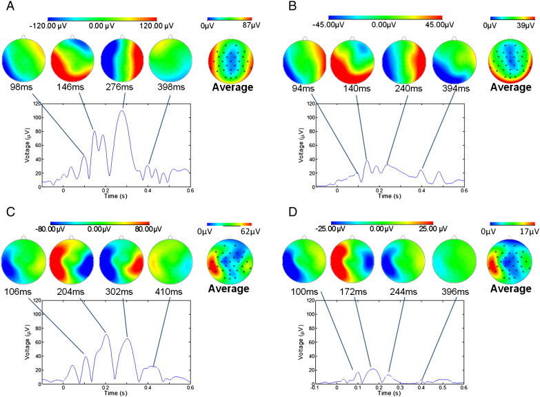
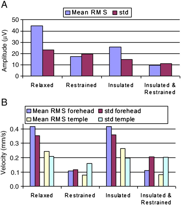
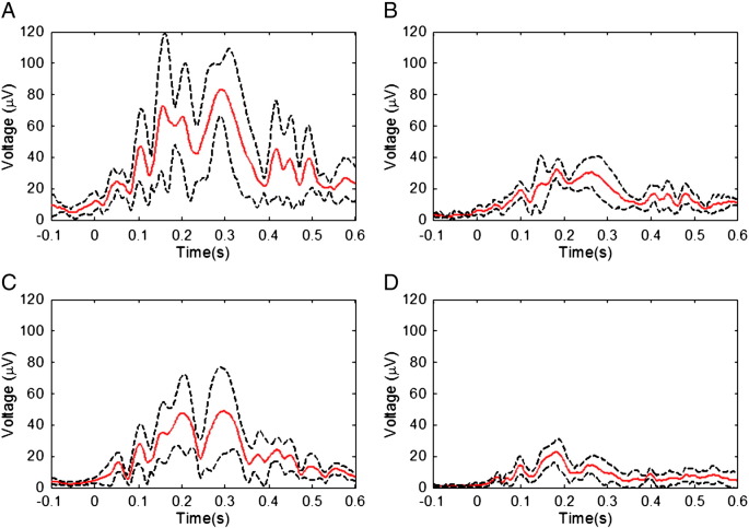
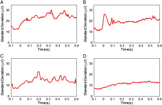
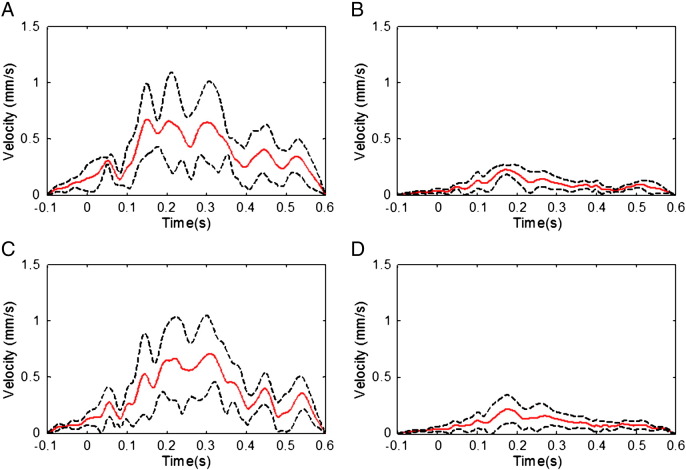
EEG recordings made during concurrent fMRI are confounded by the pulse artefact (PA), which although smaller than the gradient artefact is often more problematic because of its variability over multiple cardiac cycles. A better understanding of the PA is needed in order to generate improved methods for reducing its effect in EEG-fMRI experiments. Here we performed a study aimed at identifying the relative contributions of three putative sources of the PA (cardiac-pulse-driven head rotation, the Hall effect due to pulsatile blood flow and pulse-driven expansion of the scalp) to its amplitude and variability. EEG recordings were made from 6 subjects lying in a 3T scanner. Accelerometers were fixed on the forehead and temple to monitor head motion. A bite-bar and vacuum cushion were used to restrain the head, thus greatly attenuating the contribution of cardiac-driven head rotation to the PA, while an insulating layer placed between the head and the EEG electrodes was used to eliminate the Hall voltage contribution. Using the root mean square (RMS) amplitude of the PA averaged over leads and time as a measure of the PA amplitude, we found that head restraint and insulating layer reduced the PA by 61% and 42%, respectively, when compared with the PA induced with the subject relaxed, indicating that cardiac-pulse-driven head rotation is the dominant source of the PA. With both the insulating layer and head restraint in place, the PA was reduced in RMS amplitude by 78% compared with the relaxed condition, the remaining PA contribution resulting from scalp expansion or residual head motion. The variance of the PA across cardiac cycles was more strongly reduced by the insulating layer than the head restraint, indicating that the flow-induced Hall voltage makes a larger contribution than pulse-driven head rotation to the variability of the PA.
EEG 记录在同时进行的 fMRI 中受到脉冲伪影(PA)的干扰,尽管其幅度小于梯度伪影,但由于其在多个心动周期中的变化,通常更成问题。为了生成减少 EEG-fMRI 实验中 PA 影响的改进方法,需要更好地了解 PA。在这里,我们进行了一项研究,旨在确定 PA 的三个假定来源(由心脏脉冲驱动的头部旋转、由于脉动血流引起的 Hall 效应以及脉冲驱动的头皮扩张)对其幅度和可变性的相对贡献。我们对 6 名躺在 3T 扫描仪中的受试者进行了 EEG 记录。加速度计固定在前额和太阳穴上以监测头部运动。使用咬嘴和真空垫来限制头部,从而大大减少了心脏驱动的头部旋转对 PA 的贡献,同时在头部和 EEG 电极之间放置绝缘层以消除 Hall 电压的贡献。使用平均导联和时间的 PA 的均方根(RMS)幅度作为 PA 幅度的度量,我们发现与受试者放松时相比,头部约束和绝缘层分别将 PA 降低了 61%和 42%,表明心脏脉冲驱动的头部旋转是 PA 的主要来源。当使用绝缘层和头部约束时,与放松状态相比,PA 的 RMS 幅度降低了 78%,剩下的 PA 贡献来自头皮扩张或残余头部运动。PA 在心动周期之间的方差通过绝缘层的降低比头部约束的降低更为强烈,这表明流动诱导的 Hall 电压对 PA 的可变性的贡献大于脉冲驱动的头部旋转。