• 文献检索
  • 文档翻译
  • 深度研究
  • 学术资讯
  • Suppr Zotero 插件Zotero 插件
  • 邀请有礼
  • 套餐&价格
  • 历史记录
应用&插件
Suppr Zotero 插件Zotero 插件浏览器插件Mac 客户端Windows 客户端微信小程序
定价
高级版会员购买积分包购买API积分包
服务
文献检索文档翻译深度研究API 文档MCP 服务
关于我们
关于 Suppr公司介绍联系我们用户协议隐私条款
关注我们

Suppr 超能文献

核心技术专利:CN118964589B侵权必究
粤ICP备2023148730 号-1Suppr @ 2026

文献检索

告别复杂PubMed语法,用中文像聊天一样搜索,搜遍4000万医学文献。AI智能推荐,让科研检索更轻松。

立即免费搜索

文件翻译

保留排版,准确专业,支持PDF/Word/PPT等文件格式,支持 12+语言互译。

免费翻译文档

深度研究

AI帮你快速写综述,25分钟生成高质量综述,智能提取关键信息,辅助科研写作。

立即免费体验

用于放射治疗临床应用中的可变形图像配准验证的框架。

A framework for deformable image registration validation in radiotherapy clinical applications.

机构信息

Minneapolis Radiation Oncology, Minneapolis, MN, USA.

出版信息

J Appl Clin Med Phys. 2013 Jan 2;14(1):4066. doi: 10.1120/jacmp.v14i1.4066.

DOI:10.1120/jacmp.v14i1.4066
PMID:23318394
原文链接:https://pmc.ncbi.nlm.nih.gov/articles/PMC3732001/
Abstract

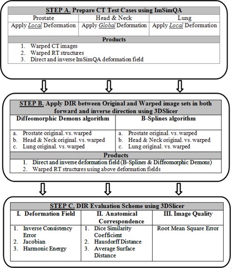
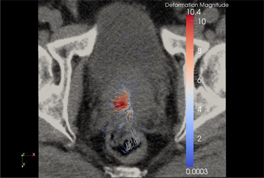
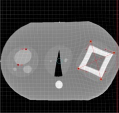
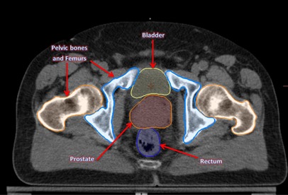
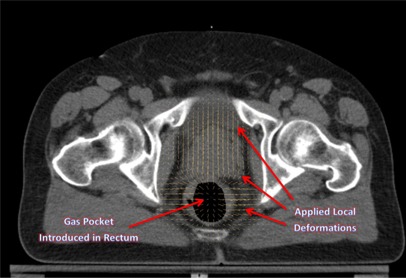
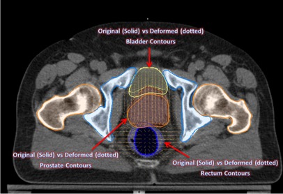
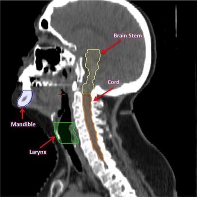
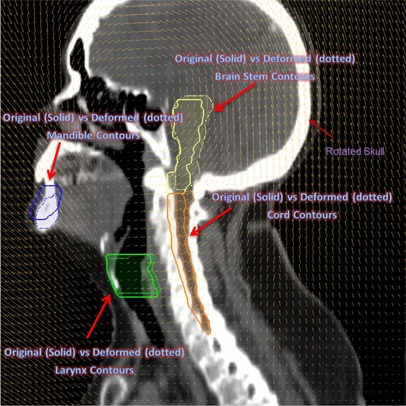
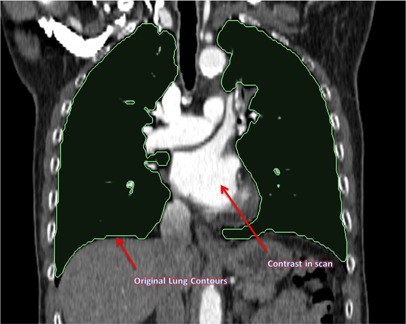
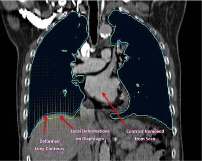
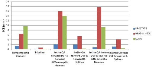
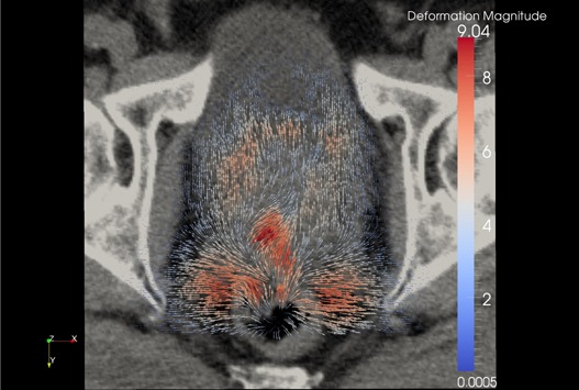
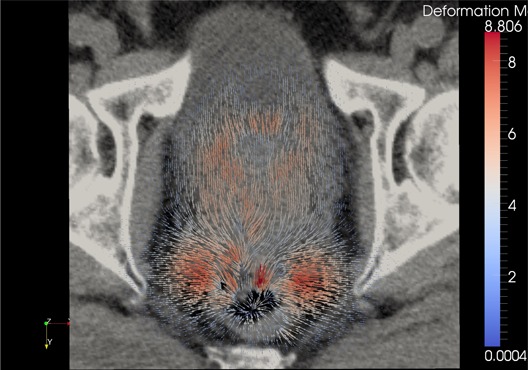
Quantitative validation of deformable image registration (DIR) algorithms is extremely difficult because of the complexity involved in constructing a deformable phantom that can duplicate various clinical scenarios. The purpose of this study is to describe a framework to test the accuracy of DIR based on computational modeling and evaluating using inverse consistency and other methods. Three clinically relevant organ deformations were created in prostate (distended rectum and rectal gas), head and neck (large neck flexion), and lung (inhale and exhale lung volumes with variable contrast enhancement) study sets. DIR was performed using both B-spline and diffeomorphic demons algorithms in the forward and inverse direction. A compositive accumulation of forward and inverse deformation vector fields was done to quantify the inverse consistency error (ICE). The anatomical correspondence of tumor and organs at risk was quantified by comparing the original RT structures with those obtained after DIR. Further, the physical characteristics of the deformation field, namely the Jacobian and harmonic energy, were computed to quantify the preservation of image topology and regularity of spatial transformation obtained in DIR. The ICE was comparable in prostate case but the B-spline algorithm had significantly better anatomical correspondence for rectum and prostate than diffeomorphic demons algorithm. The ICE was 6.5 mm for demons algorithm for head and neck case when compared to 0.7 mm for B-spline. Since the induced neck flexion was large, the average Dice similarity coefficient between both algorithms was only 0.87, 0.52, 0.81, and 0.67 for tumor, cord, parotids, and mandible, respectively. The B-spline algorithm accurately estimated deformations between images with variable contrast in our lung study, while diffeomorphic demons algorithm led to gross errors on structures affected by contrast variation. The proposed framework offers the application of known deformations on any image datasets, to evaluate the overall accuracy and limitations of a DIR algorithm used in radiation oncology. The evaluation based on anatomical correspondence, physical characteristics of deformation field, and image characteristics can facilitate DIR verification with the ultimate goal of implementing adaptive radiotherapy. The suitability of application of a particular evaluation metric in validating DIR is dependent on the clinical deformation observed.

摘要

定量验证形变图像配准(DIR)算法极其困难,因为构建能够复制各种临床场景的可变形体模非常复杂。本研究旨在描述一种基于计算建模测试 DIR 准确性的框架,并使用反向一致性和其他方法进行评估。在前列腺(扩张的直肠和直肠气体)、头颈部(大颈部弯曲)和肺部(吸气和呼气时具有不同对比增强的肺容积)研究集中创建了三个具有临床相关性的器官变形。在正向和反向方向上使用 B 样条和非刚性 demons 算法进行 DIR。对正向和反向变形矢量场进行综合积累,以量化反向一致性误差(ICE)。通过将原始 RT 结构与 DIR 后获得的结构进行比较,量化肿瘤和危险器官的解剖对应关系。此外,还计算了变形场的物理特性,即雅可比行列式和调和能量,以量化 DIR 中获得的图像拓扑和空间变换规则性的保持。在前列腺病例中,ICE 是可比的,但 B 样条算法在直肠和前列腺方面的解剖对应关系明显优于非刚性 demons 算法。在头颈部病例中,demons 算法的 ICE 为 6.5mm,而 B 样条算法的 ICE 为 0.7mm。由于诱导的颈部弯曲较大,两种算法之间的平均 Dice 相似系数分别为肿瘤、脊髓、腮腺和下颌骨的 0.87、0.52、0.81 和 0.67。B 样条算法准确估计了我们肺部研究中具有不同对比度的图像之间的变形,而非刚性 demons 算法导致受对比度变化影响的结构出现严重误差。所提出的框架提供了在任何图像数据集上应用已知变形的应用,以评估在放射肿瘤学中使用的 DIR 算法的整体准确性和局限性。基于解剖对应关系、变形场物理特性和图像特性的评估可以促进 DIR 验证,最终目标是实现自适应放疗。特定评估指标在验证 DIR 中的适用性取决于观察到的临床变形。

https://cdn.ncbi.nlm.nih.gov/pmc/blobs/1204/5714049/1f7dd80818c1/ACM2-14-192-g015.jpg
https://cdn.ncbi.nlm.nih.gov/pmc/blobs/1204/5714049/e8dfd8145fe8/ACM2-14-192-g001.jpg
https://cdn.ncbi.nlm.nih.gov/pmc/blobs/1204/5714049/372f574faab9/ACM2-14-192-g002.jpg
https://cdn.ncbi.nlm.nih.gov/pmc/blobs/1204/5714049/49892fd3c330/ACM2-14-192-g003.jpg
https://cdn.ncbi.nlm.nih.gov/pmc/blobs/1204/5714049/4718edd7f976/ACM2-14-192-g004.jpg
https://cdn.ncbi.nlm.nih.gov/pmc/blobs/1204/5714049/16323a97b4a6/ACM2-14-192-g005.jpg
https://cdn.ncbi.nlm.nih.gov/pmc/blobs/1204/5714049/baa366db5693/ACM2-14-192-g006.jpg
https://cdn.ncbi.nlm.nih.gov/pmc/blobs/1204/5714049/c66ee425dd18/ACM2-14-192-g007.jpg
https://cdn.ncbi.nlm.nih.gov/pmc/blobs/1204/5714049/84da99cb652c/ACM2-14-192-g008.jpg
https://cdn.ncbi.nlm.nih.gov/pmc/blobs/1204/5714049/d96067f8222c/ACM2-14-192-g009.jpg
https://cdn.ncbi.nlm.nih.gov/pmc/blobs/1204/5714049/c7a4f39c5424/ACM2-14-192-g010.jpg
https://cdn.ncbi.nlm.nih.gov/pmc/blobs/1204/5714049/7ef59094f94f/ACM2-14-192-g011.jpg
https://cdn.ncbi.nlm.nih.gov/pmc/blobs/1204/5714049/ecc481f60318/ACM2-14-192-g012.jpg
https://cdn.ncbi.nlm.nih.gov/pmc/blobs/1204/5714049/f4edb988aa92/ACM2-14-192-g013.jpg
https://cdn.ncbi.nlm.nih.gov/pmc/blobs/1204/5714049/d9e23feb8b41/ACM2-14-192-g014.jpg
https://cdn.ncbi.nlm.nih.gov/pmc/blobs/1204/5714049/1f7dd80818c1/ACM2-14-192-g015.jpg

相似文献

1
A framework for deformable image registration validation in radiotherapy clinical applications.用于放射治疗临床应用中的可变形图像配准验证的框架。
J Appl Clin Med Phys. 2013 Jan 2;14(1):4066. doi: 10.1120/jacmp.v14i1.4066.
2
Site-specific deformable imaging registration algorithm selection using patient-based simulated deformations.基于患者模拟变形的特定部位可变形成像配准算法选择。
Med Phys. 2013 Apr;40(4):041911. doi: 10.1118/1.4793723.
3
On the dosimetric effect and reduction of inverse consistency and transitivity errors in deformable image registration for dose accumulation.在剂量累加的形变图像配准中,针对逆一致性和传递性误差的剂量学效应及降低方法。
Med Phys. 2012 Jan;39(1):272-80. doi: 10.1118/1.3666948.
4
Analysis of deformable image registration accuracy using computational modeling.利用计算建模分析可变形图像配准的准确性。
Med Phys. 2010 Mar;37(3):970-9. doi: 10.1118/1.3302141.
5
Validation of three deformable image registration algorithms for the thorax.验证三种用于胸部的形变图像配准算法。
J Appl Clin Med Phys. 2013 Jan 7;14(1):3834. doi: 10.1120/jacmp.v14i1.3834.
6
Evaluation of various deformable image registration algorithms for thoracic images.用于胸部图像的各种可变形图像配准算法的评估。
J Radiat Res. 2014 Jan 1;55(1):175-82. doi: 10.1093/jrr/rrt093. Epub 2013 Jul 17.
7
A pseudoinverse deformation vector field generator and its applications.伪逆变形向量场生成器及其应用。
Med Phys. 2010 Mar;37(3):1117-28. doi: 10.1118/1.3301594.
8
Quantitative evaluation of a cone-beam computed tomography-planning computed tomography deformable image registration method for adaptive radiation therapy.用于自适应放射治疗的锥束计算机断层扫描-计划计算机断层扫描可变形图像配准方法的定量评估
J Appl Clin Med Phys. 2007 Nov 5;8(4):96-113. doi: 10.1120/jacmp.v8i4.2432.
9
Demons deformable registration for CBCT-guided procedures in the head and neck: convergence and accuracy.头颈部锥形束 CT 引导手术中的可变形配准:收敛性和准确性。
Med Phys. 2009 Oct;36(10):4755-64. doi: 10.1118/1.3223631.
10
The evaluation of a hybrid biomechanical deformable registration method on a multistage physical phantom with reproducible deformation.一种混合生物力学可变形配准方法在具有可重复变形的多阶段物理体模上的评估。
Radiat Oncol. 2018 Dec 4;13(1):240. doi: 10.1186/s13014-018-1192-x.

引用本文的文献

1
A viscous fluid model for large-scale motion estimation in image-guided radiotherapy.一种用于图像引导放射治疗中大规模运动估计的粘性流体模型。
Med Phys. 2025 Jul;52(7):e17967. doi: 10.1002/mp.17967.
2
Cumulative dose assessment with transformer-based deformable image registration addition for cervical cancer patients.基于变压器的可变形图像配准加法对宫颈癌患者的累积剂量评估
J Appl Clin Med Phys. 2025 Jul;26(7):e70135. doi: 10.1002/acm2.70135.
3
A fully automated machine-learning-based workflow for radiation treatment planning in prostate cancer.

本文引用的文献

1
Cone-Beam CT with a Flat-Panel Detector: From Image Science to Image-Guided Surgery.平板探测器锥形束CT:从影像科学到影像引导手术
Nucl Instrum Methods Phys Res A. 2011 Aug 21;648(S1):S241-S250. doi: 10.1016/j.nima.2010.11.088.
2
A "rolling average" multiple adaptive planning method to compensate for target volume changes in image-guided radiotherapy of prostate cancer.一种滚动平均多自适应规划方法,用于补偿前列腺癌图像引导放疗中靶区体积变化。
J Appl Clin Med Phys. 2012 Jan 5;13(1):3697. doi: 10.1120/jacmp.v13i1.3697.
3
Evaluations of an adaptive planning technique incorporating dose feedback in image-guided radiotherapy of prostate cancer.
一种用于前列腺癌放射治疗计划的基于机器学习的全自动化工作流程。
Clin Transl Radiat Oncol. 2025 Feb 11;52:100933. doi: 10.1016/j.ctro.2025.100933. eCollection 2025 May.
4
A Vessel Bifurcation Landmark Pair Dataset for Abdominal CT Deformable Image Registration (DIR) Validation.用于腹部CT可变形图像配准(DIR)验证的血管分叉地标对数据集
ArXiv. 2025 Jan 15:arXiv:2501.09162v1.
5
Accuracy Evaluation of Dose Warping Using Deformable Image Registration in Carbon Ion Therapy.碳离子治疗中使用可变形图像配准的剂量变形精度评估
Int J Part Ther. 2024 Dec 17;15:100639. doi: 10.1016/j.ijpt.2024.100639. eCollection 2025 Mar.
6
A Systematic Review on the Use of Registration-Based Change Tracking Methods in Longitudinal Radiological Images.基于登记的变化跟踪方法在纵向放射影像中的应用系统评价
J Imaging Inform Med. 2024 Nov 22. doi: 10.1007/s10278-024-01333-1.
7
Impact Assessment of Systemic Geometric Distortion in 1.5T Magnetic Resonance Imaging Simulation through Three-dimensional Geometric Distortion Phantom on Dosimetric Accuracy for Magnetic Resonance Imaging-only Prostate Treatment Planning.通过三维几何畸变体模对仅使用磁共振成像的前列腺治疗计划的剂量准确性进行1.5T磁共振成像模拟中系统几何畸变的影响评估。
J Med Phys. 2024 Jul-Sep;49(3):356-362. doi: 10.4103/jmp.jmp_62_24. Epub 2024 Sep 21.
8
Evaluation of cumulative dose distributions from external beam radiation therapy using CT-to-CBCT deformable image registration (DIR) for cervical cancer patients.使用CT到CBCT可变形图像配准(DIR)评估宫颈癌患者外照射放疗的累积剂量分布。
J Appl Clin Med Phys. 2025 Jan;26(1):e14538. doi: 10.1002/acm2.14538. Epub 2024 Oct 4.
9
Tools and recommendations for commissioning and quality assurance of deformable image registration in radiotherapy.放射治疗中可变形图像配准的委托与质量保证工具及建议。
Phys Imaging Radiat Oncol. 2024 Sep 14;32:100647. doi: 10.1016/j.phro.2024.100647. eCollection 2024 Oct.
10
Comparison of image registration methods in patients with non-melanoma skin cancer treated with superficial brachytherapy.浅表近距离放射治疗非黑色素瘤皮肤癌患者的图像配准方法比较
Phys Imaging Radiat Oncol. 2024 Aug 17;31:100631. doi: 10.1016/j.phro.2024.100631. eCollection 2024 Jul.
评估一种在图像引导前列腺癌放射治疗中结合剂量反馈的自适应计划技术。
Med Phys. 2011 Dec;38(12):6362-70. doi: 10.1118/1.3658567.
4
Adaptive radiotherapy for head-and-neck cancer: initial clinical outcomes from a prospective trial.头颈部癌症的自适应放疗:前瞻性试验的初步临床结果。
Int J Radiat Oncol Biol Phys. 2012 Jul 1;83(3):986-93. doi: 10.1016/j.ijrobp.2011.08.017. Epub 2011 Dec 2.
5
Image-guided robotic surgery: update on research and potential applications in urologic surgery.图像引导机器人手术:泌尿科手术中研究与潜在应用的最新进展。
Curr Opin Urol. 2012 Jan;22(1):47-54. doi: 10.1097/MOU.0b013e32834d4ce5.
6
A two-dimensional deformable phantom for quantitatively verifying deformation algorithms.一种用于定量验证变形算法的二维变形体模。
Med Phys. 2011 Aug;38(8):4583-6. doi: 10.1118/1.3597881.
7
Dosimetric and geometric evaluation of a hybrid strategy of offline adaptive planning and online image guidance for prostate cancer radiotherapy.前列腺癌放射治疗中离线自适应计划和在线图像引导混合策略的剂量学和几何评估。
Phys Med Biol. 2011 Aug 7;56(15):5045-62. doi: 10.1088/0031-9155/56/15/024. Epub 2011 Jul 19.
8
Dose variation during hypofractionated image-guided radiotherapy for prostate cancer: planned versus delivered.前列腺癌大分割图像引导放射治疗期间的剂量变化:计划剂量与实际给予剂量对比
J Cancer Res Ther. 2011 Apr-Jun;7(2):162-7. doi: 10.4103/0973-1482.82920.
9
Demons deformable registration of CT and cone-beam CT using an iterative intensity matching approach.基于迭代强度匹配的 CT 和锥形束 CT 配准。
Med Phys. 2011 Apr;38(4):1785-98. doi: 10.1118/1.3555037.
10
Image-guided, adaptive radiotherapy of prostate cancer: toward new standards of radiotherapy practice.影像引导下的前列腺癌自适应放疗:迈向放射治疗实践的新标准。
Front Radiat Ther Oncol. 2011;43:344-368. doi: 10.1159/000322485. Epub 2011 May 20.