Discipline of Pathology, Sydney Medical School, University of Sydney, Camperdown, New South Wales, Australia.
PLoS One. 2013;8(1):e52586. doi: 10.1371/journal.pone.0052586. Epub 2013 Jan 8.
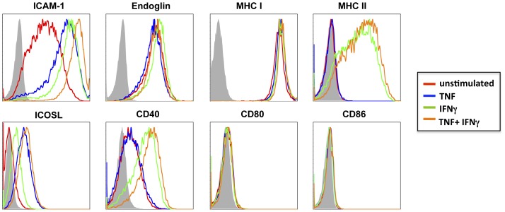
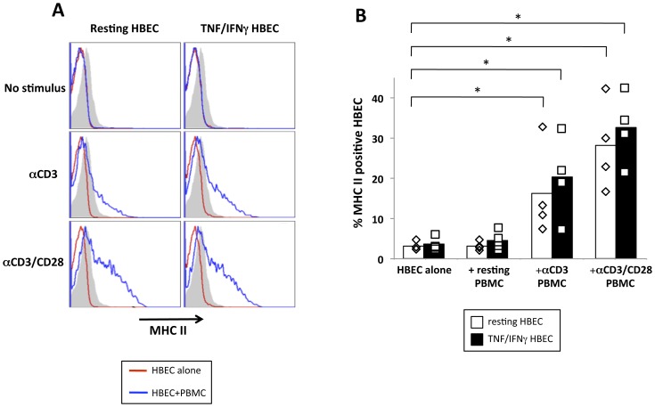
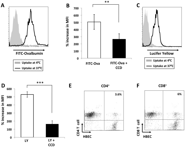
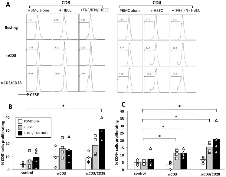
Endothelial cells (EC) form the inner lining of blood vessels and are positioned between circulating lymphocytes and tissues. Hypotheses have formed that EC may act as antigen presenting cells based on the intimate interactions with T cells, which are seen in diseases like multiple sclerosis, cerebral malaria (CM) and viral neuropathologies. Here, we investigated how human brain microvascular EC (HBEC) interact with and support the proliferation of T cells. We found HBEC to express MHC II, CD40 and ICOSL, key molecules for antigen presentation and co-stimulation and to take up fluorescently labeled antigens via macropinocytosis. In co-cultures, we showed that HBEC support and promote the proliferation of CD4(+) and CD8(+) T cells, which both are key in CM pathogenesis, particularly following T cell receptor activation and co-stimulation. Our findings provide novel evidence that HBEC can trigger T cell activation, thereby providing a novel mechanism for neuroimmunological complications of infectious diseases.
内皮细胞(EC)构成血管的内层,位于循环淋巴细胞和组织之间。基于与 T 细胞的密切相互作用,假设 EC 可能作为抗原呈递细胞发挥作用,这种相互作用在多发性硬化症、脑疟疾(CM)和病毒性神经病理学等疾病中可见。在这里,我们研究了人脑血管内皮细胞(HBEC)如何与 T 细胞相互作用并支持其增殖。我们发现 HBEC 表达 MHC II、CD40 和 ICOSL,这些都是抗原呈递和共刺激的关键分子,并通过巨胞饮作用摄取荧光标记的抗原。在共培养物中,我们表明 HBEC 支持和促进 CD4(+)和 CD8(+)T 细胞的增殖,这两者在 CM 发病机制中都很关键,特别是在 T 细胞受体激活和共刺激之后。我们的研究结果提供了新的证据,表明 HBEC 可以触发 T 细胞激活,从而为感染性疾病的神经免疫并发症提供了新的机制。