• 文献检索
  • 文档翻译
  • 深度研究
  • 学术资讯
  • Suppr Zotero 插件Zotero 插件
  • 邀请有礼
  • 套餐&价格
  • 历史记录
应用&插件
Suppr Zotero 插件Zotero 插件浏览器插件Mac 客户端Windows 客户端微信小程序
定价
高级版会员购买积分包购买API积分包
服务
文献检索文档翻译深度研究API 文档MCP 服务
关于我们
关于 Suppr公司介绍联系我们用户协议隐私条款
关注我们

Suppr 超能文献

核心技术专利:CN118964589B侵权必究
粤ICP备2023148730 号-1Suppr @ 2026

文献检索

告别复杂PubMed语法,用中文像聊天一样搜索,搜遍4000万医学文献。AI智能推荐,让科研检索更轻松。

立即免费搜索

文件翻译

保留排版,准确专业,支持PDF/Word/PPT等文件格式,支持 12+语言互译。

免费翻译文档

深度研究

AI帮你快速写综述,25分钟生成高质量综述,智能提取关键信息,辅助科研写作。

立即免费体验

一种新的基于网络的肌肉组织学自动分析方法。

A new web-based method for automated analysis of muscle histology.

机构信息

Laboratory of Molecular Myology, Friedrich-Baur-Institute, Department of Neurology, Ludwig-Maximilians-Universität München, Germany.

出版信息

BMC Musculoskelet Disord. 2013 Jan 16;14:26. doi: 10.1186/1471-2474-14-26.

DOI:10.1186/1471-2474-14-26
PMID:23324401
原文链接:https://pmc.ncbi.nlm.nih.gov/articles/PMC3560198/
Abstract

BACKGROUND

Duchenne muscular dystrophy is an inherited degenerative neuromuscular disease characterised by rapidly progressive muscle weakness. Currently, curative treatment is not available. Approaches for new treatments that improve muscle strength and quality of life depend on preclinical testing in animal models. The mdx mouse model is the most frequently used animal model for preclinical studies in muscular dystrophy research. Standardised pathology-relevant parameters of dystrophic muscle in mdx mice for histological analysis have been developed in international, collaborative efforts, but automation has not been accessible to most research groups. A standardised and mainly automated quantitative assessment of histopathological parameters in the mdx mouse model is desirable to allow an objective comparison between laboratories.

METHODS

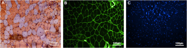
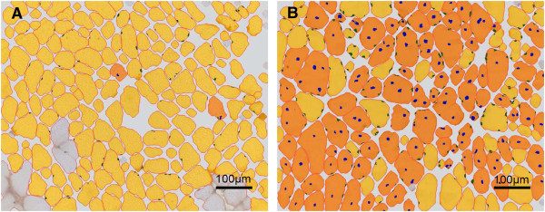
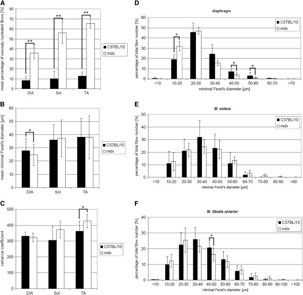
Immunological and histochemical reactions were used to obtain a double staining for fast and slow myosin. Additionally, fluorescence staining of the myofibre membranes allows defining the minimal Feret's diameter. The staining of myonuclei with the fluorescence dye bisbenzimide H was utilised to identify nuclei located internally within myofibres. Relevant structures were extracted from the image as single objects and assigned to different object classes using web-based image analysis (MyoScan). Quantitative and morphometric data were analysed, e.g. the number of nuclei per fibre and minimal Feret's diameter in 6 month old wild-type C57BL/10 mice and mdx mice.

RESULTS

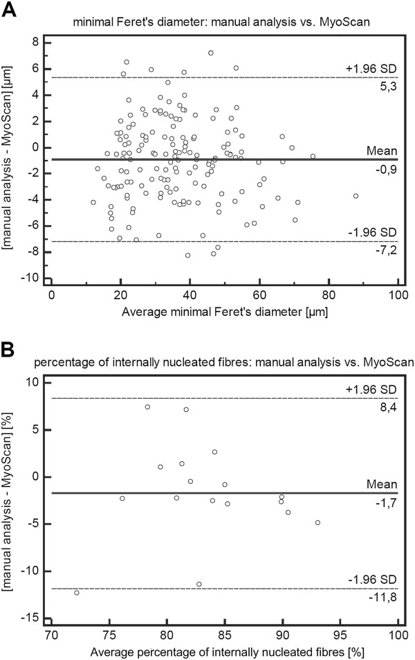
In the current version of the module "MyoScan", essential parameters for histologic analysis of muscle sections were implemented including the minimal Feret's diameter of the myofibres and the automated calculation of the percentage of internally nucleated myofibres. Morphometric data obtained in the present study were in good agreement with previously reported data in the literature and with data obtained from manual analysis.

CONCLUSIONS

A standardised and mainly automated quantitative assessment of histopathological parameters in the mdx mouse model is now available. Automated analysis of histological parameters is more rapid and less time-consuming. Moreover, results are unbiased and more reliable. Efficacy of therapeutic interventions, e.g. within the scope of a drug screening or therapeutic exon skipping, can be monitored. The automatic analysis system MyoScan used in this study is not limited exclusively to dystrophin-deficient mice but also represents a useful tool for applications in the research of other dystrophic pathologies, various other skeletal muscle diseases and degenerative neuromuscular disorders.

摘要

背景

杜氏肌营养不良症是一种遗传性进行性神经肌肉疾病,其特征是肌肉力量迅速减弱。目前,尚无治愈方法。改善肌肉力量和生活质量的新治疗方法的方法取决于在动物模型中的临床前测试。mdx 小鼠模型是肌肉营养不良症研究中最常用的临床前研究动物模型。在国际合作努力中,已经为 mdx 小鼠的肌肉病理学相关参数制定了标准化的病理学分析标准,但大多数研究小组无法实现自动化。需要对 mdx 小鼠模型中的组织病理学参数进行标准化和主要自动化评估,以允许实验室之间进行客观比较。

方法

免疫和组织化学反应用于获得快肌和慢肌的双重染色。此外,肌纤维膜的荧光染色允许定义最小 Feret 直径。利用荧光染料双苯并咪唑 H 对肌核进行染色,用于识别位于肌纤维内部的核。使用基于网络的图像分析(MyoScan)从图像中提取相关结构作为单个对象,并将它们分配到不同的对象类。分析了定量和形态计量学数据,例如 6 月龄野生型 C57BL/10 小鼠和 mdx 小鼠的每根纤维的核数和最小 Feret 直径。

结果

在当前版本的“ MyoScan”模块中,已经实现了用于肌肉切片组织学分析的基本参数,包括肌纤维的最小 Feret 直径和自动计算有核肌纤维的百分比。本研究中获得的形态计量学数据与文献中先前报道的数据以及从手动分析中获得的数据非常吻合。

结论

现在可以对 mdx 小鼠模型中的组织病理学参数进行标准化和主要自动化评估。组织学参数的自动分析更快,耗时更少。此外,结果更客观,更可靠。可以监测治疗干预的效果,例如在药物筛选或治疗性外显子跳跃的范围内。在这项研究中使用的自动分析系统 MyoScan 不仅限于肌营养不良蛋白缺乏的小鼠,而且还代表了在其他肌肉病理、各种其他骨骼肌疾病和退行性神经肌肉疾病研究中的有用工具。

https://cdn.ncbi.nlm.nih.gov/pmc/blobs/2d08/3560198/9a9d327b9ea1/1471-2474-14-26-4.jpg
https://cdn.ncbi.nlm.nih.gov/pmc/blobs/2d08/3560198/34ccf13f097a/1471-2474-14-26-1.jpg
https://cdn.ncbi.nlm.nih.gov/pmc/blobs/2d08/3560198/4ada94469bf7/1471-2474-14-26-2.jpg
https://cdn.ncbi.nlm.nih.gov/pmc/blobs/2d08/3560198/cbb0de15820b/1471-2474-14-26-3.jpg
https://cdn.ncbi.nlm.nih.gov/pmc/blobs/2d08/3560198/9a9d327b9ea1/1471-2474-14-26-4.jpg

相似文献

1
A new web-based method for automated analysis of muscle histology.一种新的基于网络的肌肉组织学自动分析方法。
BMC Musculoskelet Disord. 2013 Jan 16;14:26. doi: 10.1186/1471-2474-14-26.
2
Histological parameters for the quantitative assessment of muscular dystrophy in the mdx-mouse.用于定量评估mdx小鼠肌肉营养不良的组织学参数。
Neuromuscul Disord. 2004 Oct;14(10):675-82. doi: 10.1016/j.nmd.2004.06.008.
3
Differential expression of myosin heavy chain isoforms in the masticatory muscles of dystrophin-deficient mice.肌球蛋白重链同工型在营养不良蛋白缺陷小鼠咀嚼肌中的差异表达。
Eur J Orthod. 2011 Dec;33(6):613-9. doi: 10.1093/ejo/cjq113. Epub 2010 Dec 27.
4
Restoration of pharyngeal dilator muscle force in dystrophin-deficient (mdx) mice following co-treatment with neutralizing interleukin-6 receptor antibodies and urocortin 2.在用中和性白细胞介素-6受体抗体和尿皮质素2联合治疗后,肌营养不良蛋白缺乏(mdx)小鼠咽扩张肌力量的恢复。
Exp Physiol. 2017 Sep 1;102(9):1177-1193. doi: 10.1113/EP086232. Epub 2017 Aug 6.
5
The X-linked Becker muscular dystrophy (bmx) mouse models Becker muscular dystrophy via deletion of murine dystrophin exons 45-47.X 连锁的贝克型肌营养不良症(bmx)小鼠通过删除肌营养不良蛋白外显子 45-47 来模拟贝克型肌营养不良症。
J Cachexia Sarcopenia Muscle. 2023 Apr;14(2):940-954. doi: 10.1002/jcsm.13171. Epub 2023 Jan 11.
6
Intrinsic Muscle Stem Cell Dysfunction Contributes to Impaired Regeneration in the mdx Mouse.内在肌肉干细胞功能障碍导致mdx小鼠再生受损。
J Cachexia Sarcopenia Muscle. 2025 Feb;16(1):e13682. doi: 10.1002/jcsm.13682.
7
Correlation of Utrophin Levels with the Dystrophin Protein Complex and Muscle Fibre Regeneration in Duchenne and Becker Muscular Dystrophy Muscle Biopsies.杜兴氏和贝克氏肌营养不良症肌肉活检中肌养蛋白水平与抗肌萎缩蛋白复合体及肌纤维再生的相关性
PLoS One. 2016 Mar 14;11(3):e0150818. doi: 10.1371/journal.pone.0150818. eCollection 2016.
8
Myonucleus-related properties in soleus muscle fibers of mdx mice.mdx 小鼠比目鱼肌纤维中的核相关特性。
Cells Tissues Organs. 2010;191(3):248-59. doi: 10.1159/000240245. Epub 2009 Sep 18.
9
Distribution of MRI-derived T2 values as a biomarker for in vivo rapid screening of phenotype severity in mdx mice.作为一种生物标志物,基于 MRI 的 T2 值分布可用于体内快速筛选 mdx 小鼠的表型严重程度。
PLoS One. 2024 Sep 19;19(9):e0310551. doi: 10.1371/journal.pone.0310551. eCollection 2024.
10
Treatment With Full-Spectrum Cannabidiol Oil Improved the Pathological Findings of Dystrophic Mutant Mice.全谱大麻二酚油治疗改善了营养不良突变小鼠的病理结果。
Muscle Nerve. 2025 Apr;71(4):651-661. doi: 10.1002/mus.28337. Epub 2025 Jan 22.

引用本文的文献

1
Two Novel Mouse Models of Duchenne Muscular Dystrophy with Similar Dmd Exon 51 Frameshift Mutations and Varied Phenotype Severity.两种具有相似Dmd外显子51移码突变和不同表型严重程度的杜氏肌营养不良症新型小鼠模型。
Int J Mol Sci. 2024 Dec 27;26(1):158. doi: 10.3390/ijms26010158.
2
In Vivo Raman Spectroscopy of Muscle Is Highly Sensitive for Detection of Healthy Muscle and Highly Specific for Detection of Disease.体内拉曼光谱对肌肉具有高度敏感性,可用于检测健康肌肉,且具有高度特异性,可用于检测疾病。
Anal Chem. 2024 Oct 8;96(40):15991-15997. doi: 10.1021/acs.analchem.4c03430. Epub 2024 Sep 26.
3
Differences in meat quality between Angus cattle and Xinjiang brown cattle in association with gut microbiota and its lipid metabolism.

本文引用的文献

1
NIH Image to ImageJ: 25 years of image analysis.NIH 图像到 ImageJ:25 年的图像分析。
Nat Methods. 2012 Jul;9(7):671-5. doi: 10.1038/nmeth.2089.
2
Evaluation of the therapeutic potential of carbonic anhydrase inhibitors in two animal models of dystrophin deficient muscular dystrophy.评估碳酸酐酶抑制剂在两种肌营养不良症动物模型中的治疗潜力。
Hum Mol Genet. 2009 Nov 1;18(21):4089-101. doi: 10.1093/hmg/ddp358. Epub 2009 Jul 31.
3
Preclinical drug trials in the mdx mouse: assessment of reliable and sensitive outcome measures.
安格斯牛与新疆褐牛之间肉质差异及其与肠道微生物群及其脂质代谢的关系。
Front Microbiol. 2022 Dec 6;13:988984. doi: 10.3389/fmicb.2022.988984. eCollection 2022.
4
A User-Friendly Approach for Routine Histopathological and Morphometric Analysis of Skeletal Muscle Using CellProfiler Software.一种使用CellProfiler软件对骨骼肌进行常规组织病理学和形态计量分析的用户友好方法。
Diagnostics (Basel). 2022 Feb 22;12(3):561. doi: 10.3390/diagnostics12030561.
5
CD38-NADase is a new major contributor to Duchenne muscular dystrophic phenotype.CD38-NADase 是杜氏肌营养不良表型的一个新的主要贡献因子。
EMBO Mol Med. 2022 May 9;14(5):e12860. doi: 10.15252/emmm.202012860. Epub 2022 Mar 17.
6
Comparison of Shifts in Skeletal Muscle Plasticity Parameters in Horses in Three Different Muscles, in Answer to 8 Weeks of Harness Training.对马的三种不同肌肉中骨骼肌可塑性参数变化的比较,作为对8周挽具训练的反应。
Front Vet Sci. 2021 Oct 18;8:718866. doi: 10.3389/fvets.2021.718866. eCollection 2021.
7
Automated image segmentation method to analyse skeletal muscle cross section in exercise-induced regenerating myofibers.自动化图像分割方法分析运动诱导再生肌纤维的骨骼肌横截面积。
Sci Rep. 2021 Oct 29;11(1):21327. doi: 10.1038/s41598-021-00886-3.
8
The emerging role of the sympathetic nervous system in skeletal muscle motor innervation and sarcopenia.交感神经系统在骨骼肌运动神经支配和肌肉减少症中的新作用。
Ageing Res Rev. 2021 May;67:101305. doi: 10.1016/j.arr.2021.101305. Epub 2021 Feb 18.
9
Lymphangiogenic therapy prevents cardiac dysfunction by ameliorating inflammation and hypertension.淋巴管生成治疗通过改善炎症和高血压来预防心脏功能障碍。
Elife. 2020 Nov 17;9:e58376. doi: 10.7554/eLife.58376.
10
Semiautomatic morphometric analysis of skeletal muscle obtained by needle biopsy in older adults.老年人经皮骨骼肌活检获得的骨骼肌半自动形态计量分析。
Geroscience. 2020 Dec;42(6):1431-1443. doi: 10.1007/s11357-020-00266-1. Epub 2020 Sep 18.
杜兴氏肌营养不良症小鼠的临床前药物试验:可靠且敏感的结果指标评估
Muscle Nerve. 2009 May;39(5):591-602. doi: 10.1002/mus.21211.
4
Mammalian animal models for Duchenne muscular dystrophy.杜氏肌营养不良症的哺乳动物动物模型。
Neuromuscul Disord. 2009 Apr;19(4):241-9. doi: 10.1016/j.nmd.2008.11.015. Epub 2009 Feb 12.
5
Design, analysis, and interpretation of method-comparison studies.方法比较研究的设计、分析与解读
AACN Adv Crit Care. 2008 Apr-Jun;19(2):223-34. doi: 10.1097/01.AACN.0000318125.41512.a3.
6
Treatments for muscular dystrophy: increased treatment options for Duchenne and related muscular dystropies.肌肉萎缩症的治疗:杜兴氏及相关肌肉萎缩症的治疗选择增加
Gene Ther. 2008 Aug;15(15):1077-8. doi: 10.1038/gt.2008.97. Epub 2008 Jun 5.
7
Towards developing standard operating procedures for pre-clinical testing in the mdx mouse model of Duchenne muscular dystrophy.致力于制定杜氏肌营养不良症mdx小鼠模型临床前测试的标准操作程序。
Neurobiol Dis. 2008 Jul;31(1):1-19. doi: 10.1016/j.nbd.2008.03.008. Epub 2008 Apr 9.
8
Glucocorticoid corticosteroids for Duchenne muscular dystrophy.用于杜氏肌营养不良症的糖皮质激素皮质类固醇
Cochrane Database Syst Rev. 2008 Jan 23(1):CD003725. doi: 10.1002/14651858.CD003725.pub3.
9
Muscle regeneration in dystrophin-deficient mdx mice studied by gene expression profiling.通过基因表达谱分析研究肌营养不良蛋白缺陷型mdx小鼠的肌肉再生
BMC Genomics. 2005 Jul 13;6:98. doi: 10.1186/1471-2164-6-98.
10
Histological parameters for the quantitative assessment of muscular dystrophy in the mdx-mouse.用于定量评估mdx小鼠肌肉营养不良的组织学参数。
Neuromuscul Disord. 2004 Oct;14(10):675-82. doi: 10.1016/j.nmd.2004.06.008.