Department of Oral Anatomy and Physiology and TMD, School of Stomatology, Fourth Military Medical University, Xi'an, China.
PLoS One. 2013;8(1):e53312. doi: 10.1371/journal.pone.0053312. Epub 2013 Jan 11.
Cartilage degradation is a typical characteristic of arthritis. This study examined whether there was a subset of phagocytic chondrocytes that expressed the specific macrophage marker, CD163, and investigated their role in cartilage degradation.
Cartilage from the knee and temporomandibular joints of Sprague-Dawley rats was harvested. Cartilage degradation was experimentally-induced in rat temporomandibular joints, using published biomechanical dental methods. The expression levels of CD163 and inflammatory factors within cartilage, and the ability of CD163(+) chondrocytes to conduct phagocytosis were investigated. Cartilage from the knees of patients with osteoarthritis and normal cartilage from knee amputations was also investigated.
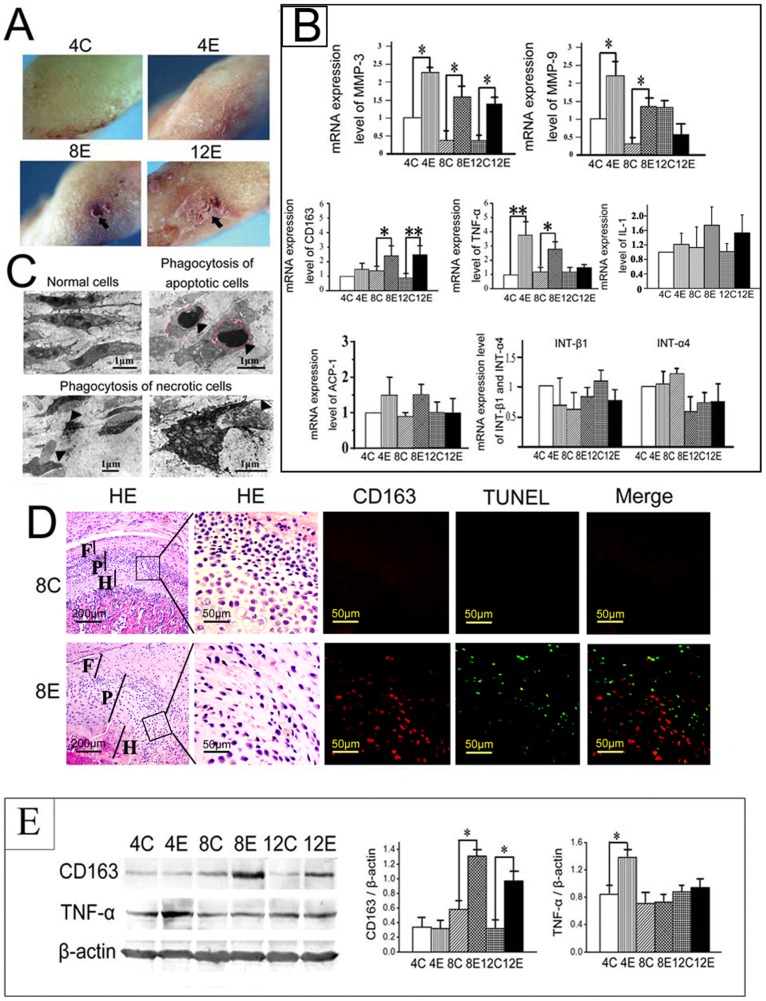
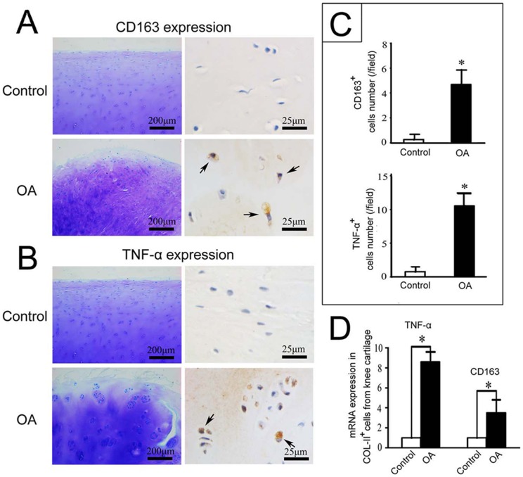
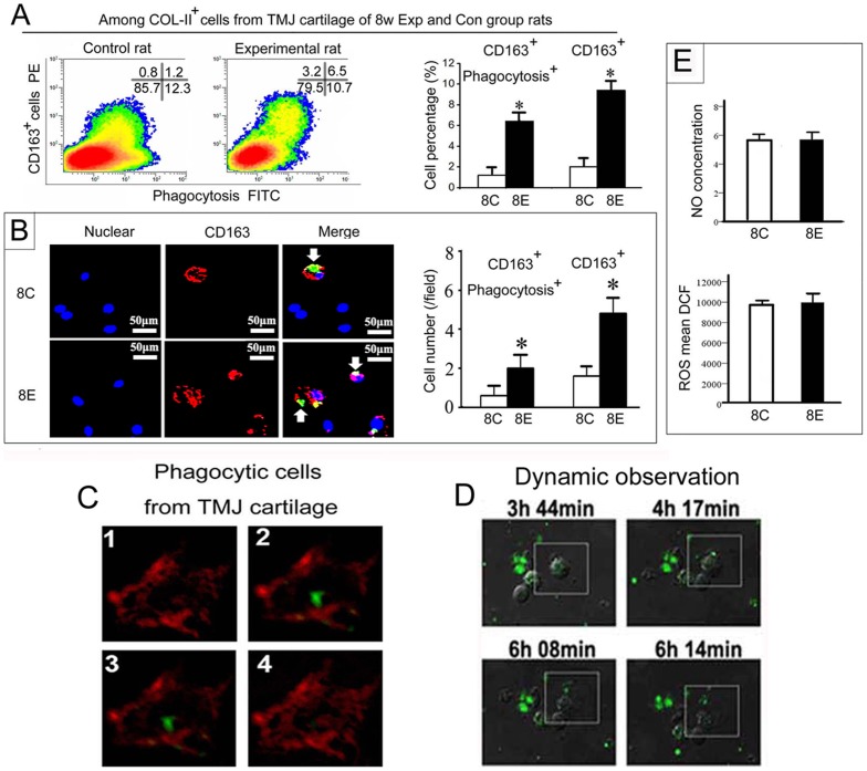
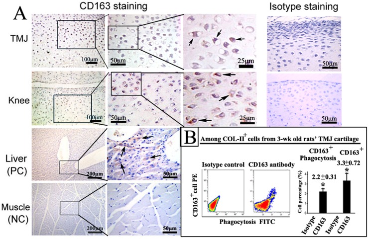
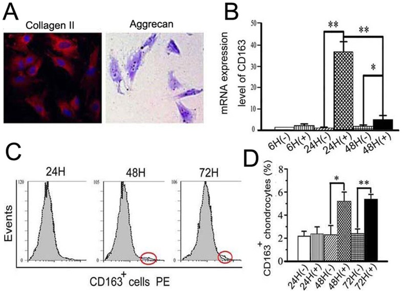
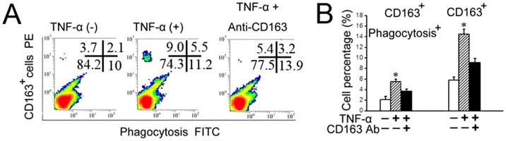
In the experimentally-induced degrading cartilage from temporomandibular joints, phagocytes were capable of engulfing neighboring apoptotic and necrotic cells, and the levels of CD163, TNF-α and MMPs were all increased (P<0.05). However, the levels of ACP-1, NO and ROS, which relate to cellular digestion capability were unchanged (P>0.05). CD163(+) chondrocytes were found in the cartilage mid-zone of temporomandibular joints and knee from healthy, three-week old rats. Furthermore, an increased number of CD163(+) chondrocytes with enhanced phagocytic activity were present in Col-II(+) chondrocytes isolated from the degraded cartilage of temporomandibular joints in the eight-week experimental group compared with their age-matched controls. Increased number with enhanced phagocytic activity of CD163(+) chondrocytes were also found in isolated Col-II(+) chondrocytes stimulated with TNF-α (P<0.05). Mid-zone distribution of CD163(+) cells accompanied with increased expression of CD163 and TNF-α were further confirmed in the isolated Col-II(+) chondrocytes from the knee cartilage of human patients with osteoarthritis, in contrast to the controls (both P<0.05).
An increased number of CD163(+) chondrocytes with enhanced phagocytic activity were discovered within degraded joint cartilage, indicating a role in eliminating degraded tissues. Targeting these cells provides a new strategy for the treatment of arthritis.
软骨降解是关节炎的一个典型特征。本研究探讨了是否存在表达特定巨噬细胞标志物 CD163 的吞噬性软骨细胞亚群,并研究了它们在软骨降解中的作用。
从 Sprague-Dawley 大鼠的膝关节和颞下颌关节中采集软骨。使用已发表的生物力学牙科方法,在大鼠颞下颌关节中实验性诱导软骨降解。研究了软骨内 CD163 和炎症因子的表达水平,以及 CD163(+)软骨细胞吞噬的能力。还研究了骨关节炎患者膝关节的软骨和膝关节截肢的正常软骨。
在实验性降解的颞下颌关节软骨中,吞噬细胞能够吞噬邻近的凋亡和坏死细胞,CD163、TNF-α 和 MMPs 的水平均升高(P<0.05)。然而,与细胞消化能力相关的 ACP-1、NO 和 ROS 水平不变(P>0.05)。在健康、三周大的大鼠的颞下颌关节和膝关节的软骨中层发现了 CD163(+)软骨细胞。此外,在 8 周实验组降解的颞下颌关节软骨中,与年龄匹配的对照组相比,Col-II(+)软骨细胞中存在更多具有增强吞噬活性的 CD163(+)软骨细胞。在 TNF-α刺激的 Col-II(+)软骨细胞中也发现了更多具有增强吞噬活性的 CD163(+)软骨细胞(P<0.05)。在骨关节炎患者膝关节软骨的 Col-II(+)软骨细胞中进一步证实了 CD163(+)细胞的中层分布以及 CD163 和 TNF-α的表达增加,与对照组相比(均 P<0.05)。
在降解的关节软骨中发现了具有增强吞噬活性的更多 CD163(+)软骨细胞,表明其在清除降解组织中起作用。针对这些细胞为关节炎的治疗提供了一种新策略。