Arthritis Res Ther. 2013;15(5):R119. doi: 10.1186/ar4299.
The repair capability of traumatized articular cartilage is highly limited so that joint injuries often lead to osteoarthritis. Migratory chondrogenic progenitor cells (CPC) might represent a target cell population for in situ regeneration. This study aims to clarify, whether 1) CPC are present in regions of macroscopically intact cartilage from human osteoarthritic joints, 2) CPC migration is stimulated by single growth factors and the cocktail of factors released from traumatized cartilage and 3) CPC migration is influenced by cytokines present in traumatized joints.
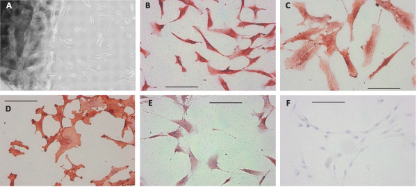
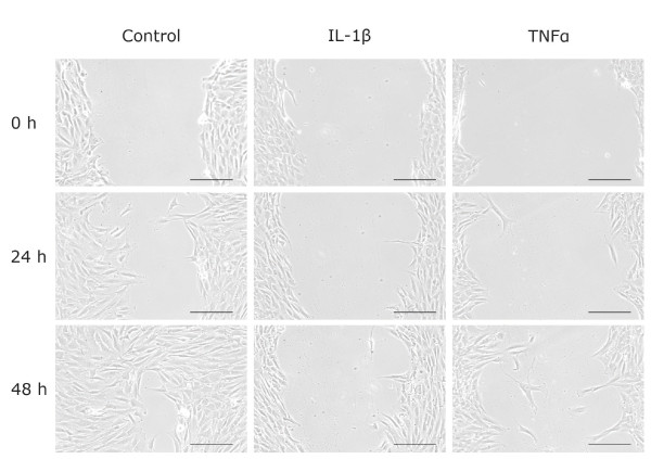
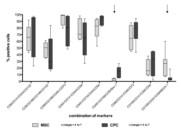
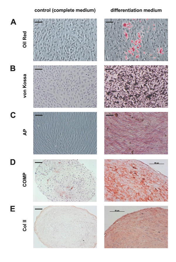
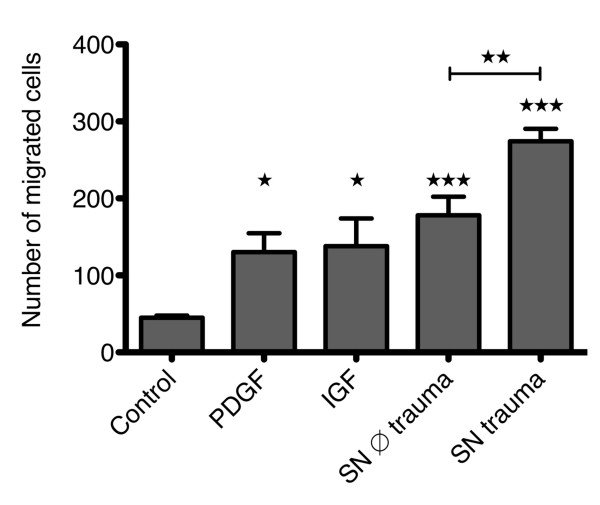
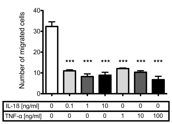
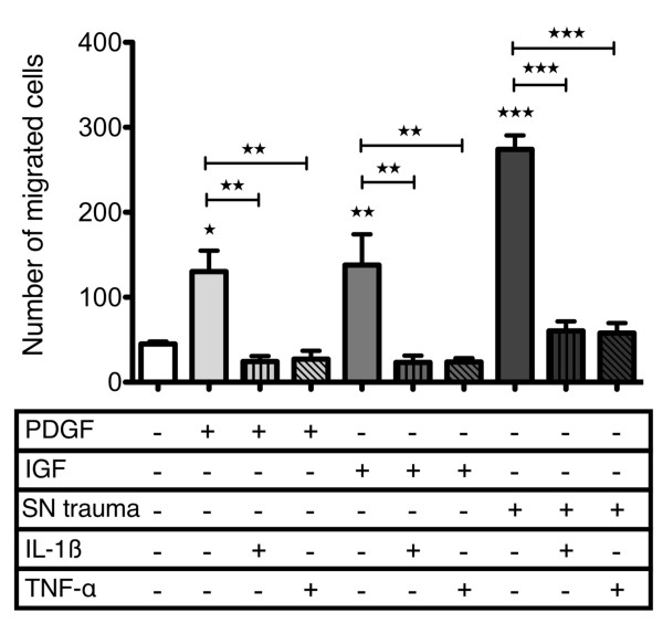
We characterized the cells growing out from macroscopically intact human osteoarthritic cartilage using a panel of positive and negative surface markers and analyzed their differentiation capacity. The migratory response to platelet-derived growth factor (PDGF)-BB, insulin-like growth factor 1 (IGF-1), supernatants obtained from in vitro traumatized cartilage and interleukin-1 beta (IL-1β) as well as tumor necrosis factor alpha (TNF-α) were tested with a modified Boyden chamber assay. The influence of IL-1β and TNF-α was additionally examined by scratch assays and outgrowth experiments.
A comparison of 25 quadruplicate marker combinations in CPC and bone-marrow derived mesenchymal stromal cells showed a similar expression profile. CPC cultures had the potential for adipogenic, osteogenic and chondrogenic differentiation. PDGF-BB and IGF-1, such as the supernatant from traumatized cartilage, induced a significant site-directed migratory response. IL-1β and TNF-α significantly reduced basal cell migration and abrogated the stimulative effect of the growth factors and the trauma supernatant. Both cytokines also inhibited cell migration in the scratch assay and primary outgrowth of CPC from cartilage tissue. In contrast, the cytokine IL-6, which is present in trauma supernatant, did not affect growth factor induced migration of CPC.
These results indicate that traumatized cartilage releases chemoattractive factors for CPC but IL-1β and TNF-α inhibit their migratory activity which might contribute to the low regenerative potential of cartilage in vivo.
创伤性关节软骨的修复能力非常有限,因此关节损伤常导致骨关节炎。迁移性软骨祖细胞(CPC)可能代表原位再生的靶细胞群。本研究旨在阐明:1)CPC 是否存在于人类骨关节炎关节的宏观完整软骨区域中;2)CPC 迁移是否受单一生长因子以及来自创伤性软骨的因子混合物的刺激;3)CPC 迁移是否受创伤关节中细胞因子的影响。
我们使用一系列阳性和阴性表面标志物对从宏观完整的人类骨关节炎软骨中生长出来的细胞进行了特征描述,并分析了它们的分化能力。使用改良 Boyden 室测定法检测血小板衍生生长因子(PDGF-BB)、胰岛素样生长因子 1(IGF-1)、体外创伤软骨获得的上清液以及白细胞介素-1β(IL-1β)和肿瘤坏死因子α(TNF-α)对迁移的反应。通过划痕试验和体外生长试验进一步研究了 IL-1β 和 TNF-α 的影响。
对 CPC 和骨髓间充质基质细胞中的 25 个四重标记组合的比较显示出相似的表达谱。CPC 培养物具有成脂、成骨和成软骨分化的潜力。PDGF-BB 和 IGF-1 以及创伤软骨的上清液诱导了明显的定向迁移反应。IL-1β 和 TNF-α 显著降低了细胞的基础迁移,并阻断了生长因子和创伤上清液的刺激作用。两种细胞因子还抑制了划痕试验中的细胞迁移和软骨组织中 CPC 的原代生长。相比之下,存在于创伤上清液中的细胞因子 IL-6 并不影响 CPC 生长因子诱导的迁移。
这些结果表明,创伤性软骨释放趋化因子吸引 CPC,但 IL-1β 和 TNF-α 抑制其迁移活性,这可能导致软骨在体内的再生潜力较低。