Department of Plastic, Reconstructive and Hand Surgery, Erasmus MC, University Medical Center, Rotterdam, The Netherlands.
PLoS One. 2013;8(1):e54041. doi: 10.1371/journal.pone.0054041. Epub 2013 Jan 10.
The purpose of this study was to determine the reliability and validity of a new non-invasive ultrasound technique to measure gastrocnemius muscle atrophy after nerve denervation in an animal model.
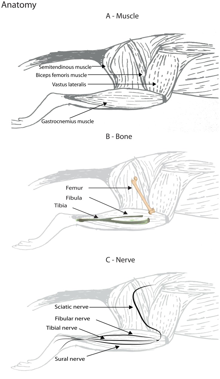
In sixteen rodents an eight mm sciatic nerve gap was created. In the following 8 weeks, each week, two rodents were euthanized and the gastrocnemius muscle was examined using two different ultrasound systems and two investigators. The standardized ultrasound measurement protocol consisted of identifying pre-defined anatomical landmarks: 1) the fibula, 2) the fibular nerve, and 3) the junction between the most distal point of the semitendinosus muscle and gastrocnemius muscle. Consequently, we measured the muscle thickness as the length of the line between the fibula and the junction between the two muscles, perpendicular to the fibular nerve. After the ultrasound recording, the muscle mass was determined.
A steep decline of muscle weight of 24% was observed after one week. In the following weeks, the weight further decreased and then remained stable from 6 weeks onwards, resulting in a maximal muscle weight decrease of 82%. The correlation coefficient was >0.96 between muscle diameter and weight using both ultrasound systems. The inter-rater reliability was excellent for both devices on the operated side (ICC of 0.99 for both ultrasound systems) and good for the non-operated site (ICC's: 0.84 & 0.89). The difference between the muscle mass ratio and the muscle thickness ratio was not more than 5% with two outliers of approximately 13%.
We have developed an innovative, highly reliable technique for quantifying muscle atrophy after nerve injury. This technique allows serial measurements in the same animal over time. This is a significant advantage compared to the conventional technique for quantifying muscle atrophy, which requires sacrificing the animal.
本研究旨在确定一种新的非侵入性超声技术在动物模型中测量神经切断后腓肠肌萎缩的可靠性和有效性。
在 16 只啮齿动物中,创建了 8 毫米的坐骨神经间隙。在接下来的 8 周内,每周有 2 只啮齿动物被安乐死,并用两种不同的超声系统和两名研究人员对腓肠肌进行检查。标准化的超声测量方案包括确定预定义的解剖学标志:1)腓骨,2)腓总神经,3)半腱肌和腓肠肌最远端之间的交界处。因此,我们测量了腓骨和两条肌肉交界处之间的直线长度,即肌肉厚度,垂直于腓总神经。超声记录后,测量肌肉质量。
术后一周,肌肉重量急剧下降 24%。在接下来的几周里,肌肉重量进一步下降,然后从 6 周开始保持稳定,导致最大肌肉重量减少 82%。使用两种超声系统,肌肉直径与重量之间的相关系数均>0.96。两种设备在手术侧的组内相关系数均为 0.99,非手术侧的组内相关系数为 0.84 和 0.89,均为优秀。肌肉质量比和肌肉厚度比之间的差异不超过 5%,有两个约为 13%的离群值。
我们开发了一种创新的、高度可靠的技术,用于量化神经损伤后的肌肉萎缩。这种技术允许在同一动物中随时间进行系列测量。与量化肌肉萎缩的传统技术相比,这是一个显著的优势,因为传统技术需要牺牲动物。