Electrical and Electronics Engineering Department, Bilkent University, Ankara, Turkey.
PLoS One. 2013;8(1):e52807. doi: 10.1371/journal.pone.0052807. Epub 2013 Jan 16.
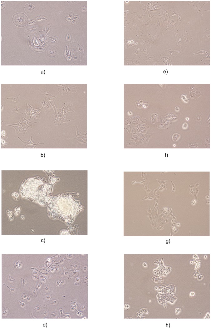
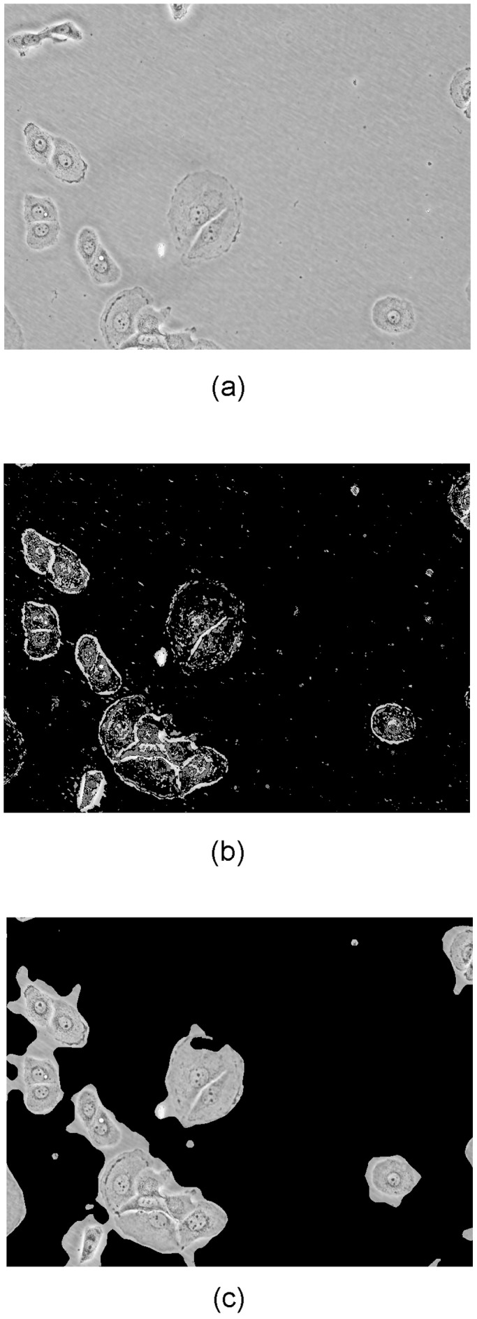
Cancer cell lines are widely used for research purposes in laboratories all over the world. Computer-assisted classification of cancer cells can alleviate the burden of manual labeling and help cancer research. In this paper, we present a novel computerized method for cancer cell line image classification. The aim is to automatically classify 14 different classes of cell lines including 7 classes of breast and 7 classes of liver cancer cells. Microscopic images containing irregular carcinoma cell patterns are represented by subwindows which correspond to foreground pixels. For each subwindow, a covariance descriptor utilizing the dual-tree complex wavelet transform (DT-[Formula: see text]WT) coefficients and several morphological attributes are computed. Directionally selective DT-[Formula: see text]WT feature parameters are preferred primarily because of their ability to characterize edges at multiple orientations which is the characteristic feature of carcinoma cell line images. A Support Vector Machine (SVM) classifier with radial basis function (RBF) kernel is employed for final classification. Over a dataset of 840 images, we achieve an accuracy above 98%, which outperforms the classical covariance-based methods. The proposed system can be used as a reliable decision maker for laboratory studies. Our tool provides an automated, time- and cost-efficient analysis of cancer cell morphology to classify different cancer cell lines using image-processing techniques, which can be used as an alternative to the costly short tandem repeat (STR) analysis. The data set used in this manuscript is available as supplementary material through http://signal.ee.bilkent.edu.tr/cancerCellLineClassificationSampleImages.html.
癌细胞系在世界各地的实验室中被广泛用于研究目的。计算机辅助的癌细胞分类可以减轻手动标记的负担,有助于癌症研究。在本文中,我们提出了一种新颖的癌细胞系图像分类计算机方法。目的是自动分类 14 种不同类型的细胞系,包括 7 种乳腺癌细胞系和 7 种肝癌细胞系。包含不规则癌细胞模式的显微镜图像由对应于前景像素的子窗口表示。对于每个子窗口,计算利用双树复小波变换 (DT-[Formula: see text]WT) 系数和几个形态属性的协方差描述符。首选具有方向选择性的 DT-[Formula: see text]WT 特征参数主要是因为它们能够在多个方向上表征边缘,这是癌细胞系图像的特征。使用具有径向基函数 (RBF) 核的支持向量机 (SVM) 分类器进行最终分类。在 840 张图像的数据集上,我们的准确率超过 98%,优于经典的基于协方差的方法。所提出的系统可以作为实验室研究的可靠决策工具。我们的工具使用图像处理技术提供了一种自动化、省时且具有成本效益的癌症细胞形态分析方法,可替代昂贵的短串联重复序列 (STR) 分析。本文使用的数据集可通过 http://signal.ee.bilkent.edu.tr/cancerCellLineClassificationSampleImages.html 作为补充材料获得。