• 文献检索
  • 文档翻译
  • 深度研究
  • 学术资讯
  • Suppr Zotero 插件Zotero 插件
  • 邀请有礼
  • 套餐&价格
  • 历史记录
应用&插件
Suppr Zotero 插件Zotero 插件浏览器插件Mac 客户端Windows 客户端微信小程序
定价
高级版会员购买积分包购买API积分包
服务
文献检索文档翻译深度研究API 文档MCP 服务
关于我们
关于 Suppr公司介绍联系我们用户协议隐私条款
关注我们

Suppr 超能文献

核心技术专利:CN118964589B侵权必究
粤ICP备2023148730 号-1Suppr @ 2026

文献检索

告别复杂PubMed语法,用中文像聊天一样搜索,搜遍4000万医学文献。AI智能推荐,让科研检索更轻松。

立即免费搜索

文件翻译

保留排版,准确专业,支持PDF/Word/PPT等文件格式,支持 12+语言互译。

免费翻译文档

深度研究

AI帮你快速写综述,25分钟生成高质量综述,智能提取关键信息,辅助科研写作。

立即免费体验

基于双树复小波变换的乳腺微钙化计算机辅助诊断。

Computer-aided diagnosis of breast microcalcifications based on dual-tree complex wavelet transform.

机构信息

College of Biomedical Engineering, Capital Medical University, Beijing 100069, People's Republic of China.

出版信息

Biomed Eng Online. 2012 Dec 19;11:96. doi: 10.1186/1475-925X-11-96.

DOI:10.1186/1475-925X-11-96
PMID:23253202
原文链接:https://pmc.ncbi.nlm.nih.gov/articles/PMC3537591/
Abstract

BACKGROUND

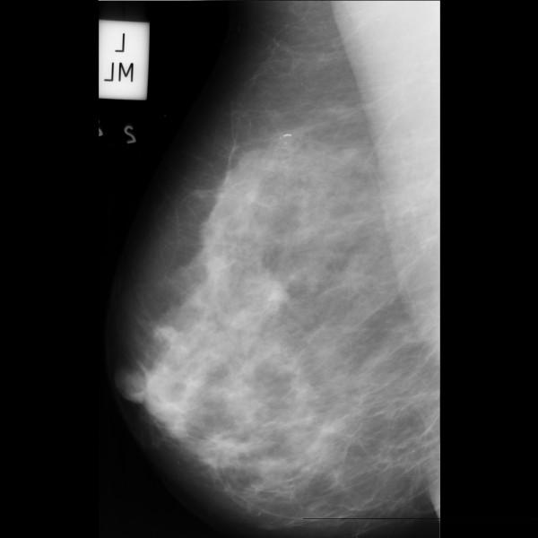
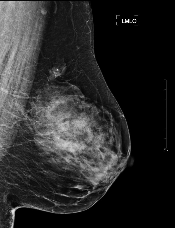
Digital mammography is the most reliable imaging modality for breast carcinoma diagnosis and breast micro-calcifications is regarded as one of the most important signs on imaging diagnosis. In this paper, a computer-aided diagnosis (CAD) system is presented for breast micro-calcifications based on dual-tree complex wavelet transform (DT-CWT) to facilitate radiologists like double reading.

METHODS

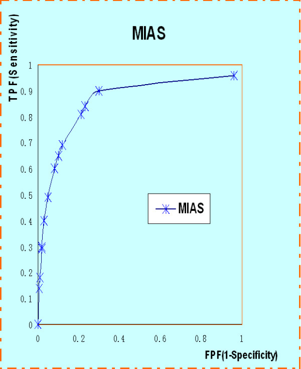
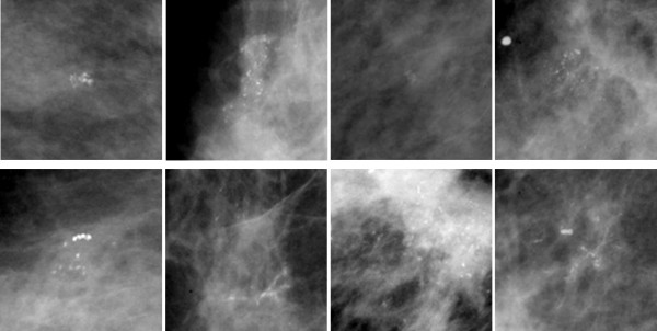
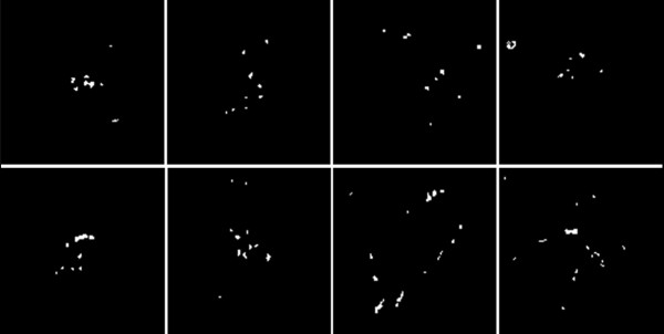
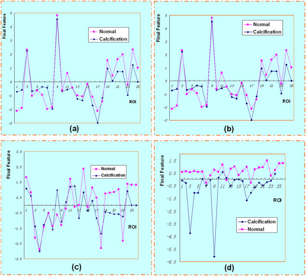
Firstly, 25 abnormal ROIs were extracted according to the center and diameter of the lesions manually and 25 normal ROIs were selected randomly. Then micro-calcifications were segmented by combining space and frequency domain techniques. We extracted three texture features based on wavelet (Haar, DB4, DT-CWT) transform. Totally 14 descriptors were introduced to define the characteristics of the suspicious micro-calcifications. Principal Component Analysis (PCA) was used to transform these descriptors to a compact and efficient vector expression. Support Vector Machine (SVM) classifier was used to classify potential micro-calcifications. Finally, we used the receiver operating characteristic (ROC) curve and free-response operating characteristic (FROC) curve to evaluate the performance of the CAD system.

RESULTS

The results of SVM classifications based on different wavelets shows DT-CWT has a better performance. Compared with other results, DT-CWT method achieved an accuracy of 96% and 100% for the classification of normal and abnormal ROIs, and the classification of benign and malignant micro-calcifications respectively. In FROC analysis, our CAD system for clinical dataset detection achieved a sensitivity of 83.5% at a false positive per image of 1.85.

CONCLUSIONS

Compared with general wavelets, DT-CWT could describe the features more effectively, and our CAD system had a competitive performance.

摘要

背景

数字乳腺摄影是乳腺癌诊断最可靠的成像方式,而乳腺微钙化被认为是影像学诊断中最重要的标志之一。在本文中,提出了一种基于双树复小波变换(DT-CWT)的乳腺微钙化计算机辅助诊断(CAD)系统,以方便放射科医生进行双重阅读。

方法

首先,根据病变的中心和直径手动提取 25 个异常 ROI,然后随机选择 25 个正常 ROI。然后,结合空域和频域技术对微钙化进行分割。我们基于小波(Haar、DB4、DT-CWT)变换提取了三个纹理特征。总共引入了 14 个描述符来定义可疑微钙化的特征。主成分分析(PCA)用于将这些描述符转换为紧凑高效的向量表示。支持向量机(SVM)分类器用于分类潜在的微钙化。最后,我们使用接收者操作特征(ROC)曲线和自由响应操作特征(FROC)曲线来评估 CAD 系统的性能。

结果

基于不同小波的 SVM 分类结果表明,DT-CWT 具有更好的性能。与其他结果相比,DT-CWT 方法对正常和异常 ROI 的分类分别达到了 96%和 100%的准确率,对良性和恶性微钙化的分类分别达到了 96%和 100%的准确率。在 FROC 分析中,我们的 CAD 系统对临床数据集的检测在每张图像的假阳性率为 1.85 时达到了 83.5%的灵敏度。

结论

与一般小波相比,DT-CWT 可以更有效地描述特征,并且我们的 CAD 系统具有竞争力的性能。

https://cdn.ncbi.nlm.nih.gov/pmc/blobs/ed53/3537591/5c640fb14030/1475-925X-11-96-7.jpg
https://cdn.ncbi.nlm.nih.gov/pmc/blobs/ed53/3537591/07c5a7f71190/1475-925X-11-96-2.jpg
https://cdn.ncbi.nlm.nih.gov/pmc/blobs/ed53/3537591/8491b70a0857/1475-925X-11-96-3.jpg
https://cdn.ncbi.nlm.nih.gov/pmc/blobs/ed53/3537591/d636efd30be2/1475-925X-11-96-4.jpg
https://cdn.ncbi.nlm.nih.gov/pmc/blobs/ed53/3537591/2cb0bad6661c/1475-925X-11-96-5.jpg
https://cdn.ncbi.nlm.nih.gov/pmc/blobs/ed53/3537591/dff1d09ae04c/1475-925X-11-96-6.jpg
https://cdn.ncbi.nlm.nih.gov/pmc/blobs/ed53/3537591/5c640fb14030/1475-925X-11-96-7.jpg

相似文献

1
Computer-aided diagnosis of breast microcalcifications based on dual-tree complex wavelet transform.基于双树复小波变换的乳腺微钙化计算机辅助诊断。
Biomed Eng Online. 2012 Dec 19;11:96. doi: 10.1186/1475-925X-11-96.
2
Breast microcalcifications detection based on fusing features with DTCWT.基于 DTCWT 融合特征的乳腺微钙化检测。
J Xray Sci Technol. 2020;28(2):197-218. doi: 10.3233/XST-190583.
3
Automated diagnosis of mammogram images of breast cancer using discrete wavelet transform and spherical wavelet transform features: a comparative study.使用离散小波变换和球面小波变换特征的乳腺癌乳房X光图像自动诊断:一项比较研究。
Technol Cancer Res Treat. 2014 Dec;13(6):605-15. doi: 10.7785/tcrtexpress.2013.600262. Epub 2013 Aug 31.
4
SVM based system for classification of microcalcifications in digital mammograms.基于支持向量机的数字乳腺X线摄影中微钙化分类系统。
Conf Proc IEEE Eng Med Biol Soc. 2006;2006:4747-50. doi: 10.1109/IEMBS.2006.259320.
5
Optimally weighted wavelet transform based on supervised training for detection of microcalcifications in digital mammograms.基于监督训练的最优加权小波变换用于数字乳腺钼靶片中微钙化的检测
Med Phys. 1998 Jun;25(6):949-56. doi: 10.1118/1.598273.
6
Computer-aided mass detection in mammography: false positive reduction via gray-scale invariant ranklet texture features.乳腺钼靶摄影中的计算机辅助肿块检测:通过灰度不变秩let纹理特征减少假阳性
Med Phys. 2009 Feb;36(2):311-6. doi: 10.1118/1.3049588.
7
Quantitative comparison of clustered microcalcifications in for-presentation and for-processing mammograms in full-field digital mammography.全数字化乳腺摄影中用于诊断和存档的钼靶片中小钙化簇的定量比较。
Med Phys. 2017 Jul;44(7):3726-3738. doi: 10.1002/mp.12316. Epub 2017 Jun 9.
8
A Hybridized ELM for Automatic Micro Calcification Detection in Mammogram Images Based on Multi-Scale Features.基于多尺度特征的乳腺钼钯图像中自动微钙化检测的混合 ELM
J Med Syst. 2019 May 15;43(7):183. doi: 10.1007/s10916-019-1316-3.
9
Evaluation of a wavelet-based computer-assisted detection system for identifying microcalcifications in digital full-field mammography.基于小波的计算机辅助检测系统在数字全视野乳腺摄影中识别微钙化的评估。
Acta Radiol. 2004 Apr;45(2):136-41. doi: 10.1080/02841850410003770.
10
Computer aided detection system for micro calcifications in digital mammograms.数字乳腺钼靶片中微钙化的计算机辅助检测系统
Comput Methods Programs Biomed. 2014 Oct;116(3):226-35. doi: 10.1016/j.cmpb.2014.04.010. Epub 2014 Apr 30.

引用本文的文献

1
Artificial Intelligence-Powered Mammography: Navigating the Landscape of Deep Learning for Breast Cancer Detection.人工智能助力的乳腺钼靶检查:探索用于乳腺癌检测的深度学习领域
Cureus. 2024 Mar 26;16(3):e56945. doi: 10.7759/cureus.56945. eCollection 2024 Mar.
2
A Hybrid Deep Transfer Learning of CNN-Based LR-PCA for Breast Lesion Diagnosis via Medical Breast Mammograms.基于 CNN 的 LR-PCA 的混合深度迁移学习在医学乳腺 X 光片中用于乳腺病变诊断。
Sensors (Basel). 2022 Jun 30;22(13):4938. doi: 10.3390/s22134938.
3
Artificial Intelligence in Medical Imaging of the Breast.

本文引用的文献

1
Computed-aided diagnosis (CAD) in the detection of breast cancer.计算机辅助诊断(CAD)在乳腺癌检测中的应用。
Eur J Radiol. 2013 Mar;82(3):417-23. doi: 10.1016/j.ejrad.2012.03.005. Epub 2012 Aug 30.
2
CADx of mammographic masses and clustered microcalcifications: a review.乳腺钼靶肿块及簇状微钙化的计算机辅助诊断:综述
Med Phys. 2009 Jun;36(6):2052-68. doi: 10.1118/1.3121511.
3
Computer-aided detection and diagnosis of breast cancer with mammography: recent advances.乳腺钼靶摄影术对乳腺癌的计算机辅助检测与诊断:最新进展
人工智能在乳腺医学成像中的应用
Front Oncol. 2021 Jul 22;11:600557. doi: 10.3389/fonc.2021.600557. eCollection 2021.
4
A machine learning approach on multiscale texture analysis for breast microcalcification diagnosis.基于多尺度纹理分析的机器学习方法在乳腺微钙化诊断中的应用。
BMC Bioinformatics. 2020 Mar 11;21(Suppl 2):91. doi: 10.1186/s12859-020-3358-4.
5
Retinal texture biomarkers may help to discriminate between Alzheimer's, Parkinson's, and healthy controls.视网膜纹理生物标志物可帮助区分阿尔茨海默病、帕金森病和健康对照者。
PLoS One. 2019 Jun 21;14(6):e0218826. doi: 10.1371/journal.pone.0218826. eCollection 2019.
6
Design and development of a content-based medical image retrieval system for spine vertebrae irregularity.用于脊椎不规则性的基于内容的医学图像检索系统的设计与开发。
Biomed Eng Online. 2015 Jan 16;14:6. doi: 10.1186/1475-925X-14-6.
IEEE Trans Inf Technol Biomed. 2009 Mar;13(2):236-51. doi: 10.1109/TITB.2008.2009441. Epub 2009 Jan 20.
4
Content-based retrieval of mammograms using visual features related to breast density patterns.利用与乳房密度模式相关的视觉特征对乳腺钼靶图像进行基于内容的检索。
J Digit Imaging. 2007 Jun;20(2):172-90. doi: 10.1007/s10278-007-9004-0. Epub 2007 Feb 22.
5
Computer-aided diagnosis scheme using a filter bank for detection of microcalcification clusters in mammograms.使用滤波器组的计算机辅助诊断方案用于检测乳腺X线照片中的微钙化簇。
IEEE Trans Biomed Eng. 2006 Feb;53(2):273-83. doi: 10.1109/TBME.2005.862536.
6
Computer-aided detection (CAD) in mammography: does it help the junior or the senior radiologist?乳腺钼靶摄影中的计算机辅助检测(CAD):它对初级放射科医生还是高级放射科医生有帮助?
Eur J Radiol. 2005 Apr;54(1):90-6. doi: 10.1016/j.ejrad.2004.11.021.
7
An automatic microcalcification detection system based on a hybrid neural network classifier.
Artif Intell Med. 2002 Jun;25(2):149-67. doi: 10.1016/s0933-3657(02)00013-1.
8
A computer-aided diagnosis system for digital mammograms based on fuzzy-neural and feature extraction techniques.
IEEE Trans Inf Technol Biomed. 2001 Mar;5(1):46-54. doi: 10.1109/4233.908389.
9
False-positive reduction in CAD mass detection using a competitive classification strategy.使用竞争分类策略减少CAD肿块检测中的假阳性
Med Phys. 2001 Feb;28(2):250-8. doi: 10.1118/1.1344203.
10
A computer-aided design mammography screening system for detection and classification of microcalcifications.一种用于微钙化检测和分类的计算机辅助设计乳腺钼靶筛查系统。
Int J Med Inform. 2000 Oct;60(1):29-57. doi: 10.1016/s1386-5056(00)00067-8.