Zhu Biyun, Luo Wei, Li Baoping, Chen Budong, Yang Qiuying, Xu Yan, Wu Xiaohua, Chen Hui, Zhang Kuan
School of Biomedical Engineering, Capital Medical University, Beijing 100069, China.
Biomed Eng Online. 2014 Oct 2;13:141. doi: 10.1186/1475-925X-13-141.
To diagnose pneumoconiosis using a computer-aided diagnosis system based on digital chest radiographs.
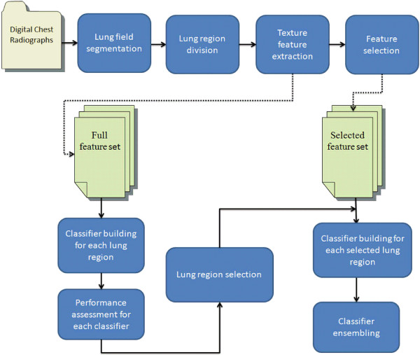
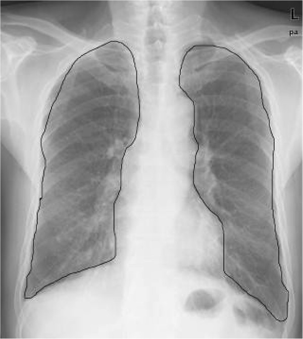
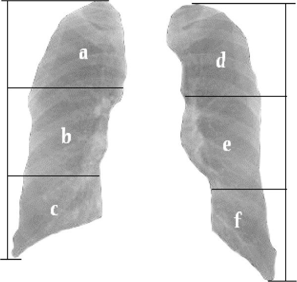
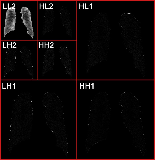
Lung fields were first extracted by combining the traditional Otsu-threshold method with a morphological reconstruction on digital radiographs (DRs), and then subdivided into six non-overlapping regions (region (a-f)). Twenty-two wavelet-based energy texture features were calculated exclusively from each region and selected using a decision tree algorithm. A support vector machine (SVM) with a linear kernel was trained using samples with texture features to classify an individual region of a healthy subject or a pneumoconiosis patient. The final classification results were obtained by integrating these individual classifiers with the weighted voting method. All models were developed on a dataset of 85 healthy controls and 40 stage I or II pneumoconiosis patients and validated by using the bootstrap resampling with replacement method.
The areas under receiver operating characteristic curves (AUCs) of regions (c) and (f) were 0.688 and 0.563, which were worse than those of the other four regions. Region (c) and (f) were both excluded from the individual classifiers that were going to be assembled further. When built on the selected texture features, each individual SVM showed a higher diagnostic performance for the training set and the test set. The classification performance after an ensemble was 0.997 and 0.961 of the AUC value for the training and test sets, respectively. The final results were 0.974 ± 0.018 for AUC value and 0.929 ± 0.018 for accuracy.
The integrated SVM model built on the selected feature set showed the highest diagnostic performance among all individual SVM models. The model has good potential in diagnosing pneumoconiosis based on digital chest radiographs.
使用基于数字化胸部X线片的计算机辅助诊断系统诊断尘肺病。
首先通过将传统的大津阈值法与数字X线片(DR)上的形态学重建相结合来提取肺野,然后将其细分为六个不重叠的区域(区域(a - f))。专门从每个区域计算22个基于小波的能量纹理特征,并使用决策树算法进行选择。使用具有纹理特征的样本训练具有线性核的支持向量机(SVM),以对健康受试者或尘肺病患者的单个区域进行分类。通过使用加权投票法整合这些单个分类器来获得最终分类结果。所有模型均基于85名健康对照和40名I期或II期尘肺病患者的数据集开发,并通过有放回的自助重采样方法进行验证。
区域(c)和(f)的受试者工作特征曲线(AUC)下面积分别为0.688和0.563,比其他四个区域差。区域(c)和(f)都被排除在将进一步组装的单个分类器之外。基于所选纹理特征构建时,每个单独的SVM对训练集和测试集均显示出更高的诊断性能。集成后的分类性能对于训练集和测试集的AUC值分别为0.997和0.961。最终结果的AUC值为0.974±0.018,准确率为0.929±0.018。
基于所选特征集构建的集成SVM模型在所有单个SVM模型中显示出最高的诊断性能。该模型在基于数字化胸部X线片诊断尘肺病方面具有良好的潜力。