Jędrych Marian, Wawryk-Gawda Ewelina, Jodłowska-Jędrych Barbara, Chylińska-Wrzos Patrycja, Jasiński Ludwik
Department of Mathematics and Biostatistics, Medical University of Lublin, 20-090, Lublin ul. K. Jaczewskiego 4, Poland.
Protoplasma. 2013 Oct;250(5):1025-34. doi: 10.1007/s00709-012-0461-z. Epub 2013 Jan 24.
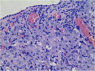
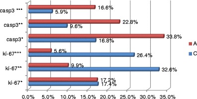
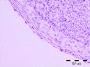
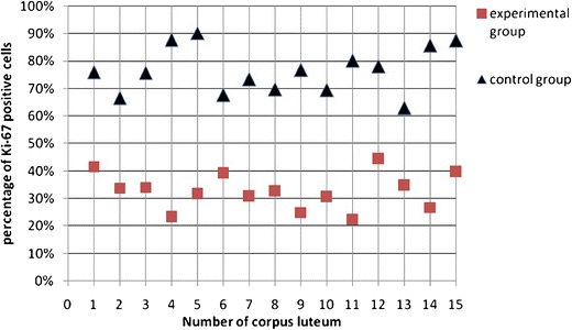
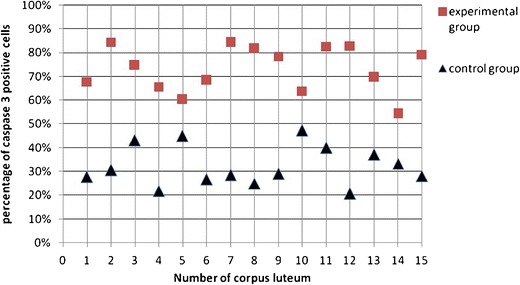
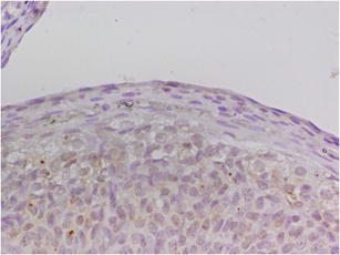
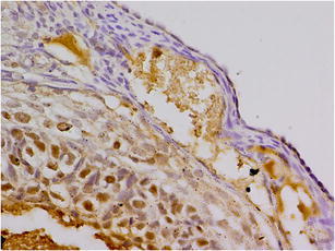
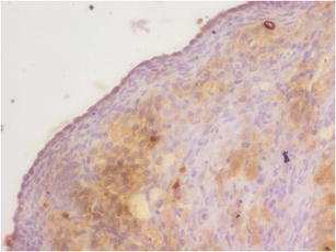
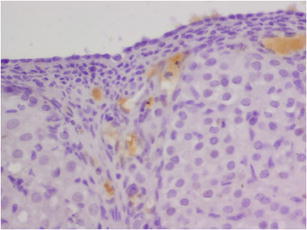
Cladribine has been used in the treatment of hairy cell leukemia for about 30 years. In addition, the number of indications for the application of 2-CdA is constantly increasing. The treatment with cladribine, of younger persons and even children, appears to be a major factor stimulating the more exact recognition of its activities. However, till now, little has been known about the impact of cladribine on the reproductive system. The aim of the study was to evaluate the immunohistochemical expression of cell proliferation and apoptosis markers in ovarian surface epithelial (OSE) cells. In our study, ten rats were placed into two equal groups. The study group received daily subcutaneous injections of cladribine in a dose of 0.10 mg/kg of weight/day for one cycle lasting 7 days. The control group received only saline injections. The rats were sacrificed 24 h after the last injection, and their ovaries were extracted. The sections were immunohistochemically stained with cell proliferation marker Ki-67 and the apoptosis marker caspase 3. The expressions of the markers were evaluated using a light microscope. An analysis was made using an image analysis system and the CellAD software. The results were then statistically explored by way of the Mann-Whitney U test. The proliferative index (Ki-67) of ovarian surface epithelial cells was significantly lower in the study group than in the control group (p < 0.05). These results suggest that cladribine treatment has a potential to inhibit the OSE cell proliferation in rats. The apoptosis marker demonstrated a significant increase after the cladribine treatment. These suggest that cladribine induces apoptosis in OSE cells.
克拉屈滨已用于治疗毛细胞白血病约30年。此外,2-氯脱氧腺苷的应用适应症数量也在不断增加。对年轻人甚至儿童使用克拉屈滨进行治疗,似乎是促使人们更准确认识其作用的一个主要因素。然而,迄今为止,关于克拉屈滨对生殖系统的影响知之甚少。本研究的目的是评估卵巢表面上皮(OSE)细胞中细胞增殖和凋亡标志物的免疫组化表达。在我们的研究中,将10只大鼠分成两个相等的组。研究组每天皮下注射剂量为0.10 mg/kg体重/天的克拉屈滨,持续一个7天的周期。对照组仅注射生理盐水。在最后一次注射后24小时处死大鼠,并取出它们的卵巢。切片用细胞增殖标志物Ki-67和凋亡标志物半胱天冬酶3进行免疫组化染色。使用光学显微镜评估标志物的表达。使用图像分析系统和CellAD软件进行分析。然后通过曼-惠特尼U检验对结果进行统计学探究。研究组卵巢表面上皮细胞的增殖指数(Ki-67)明显低于对照组(p < 0.05)。这些结果表明,克拉屈滨治疗有可能抑制大鼠OSE细胞的增殖。克拉屈滨治疗后凋亡标志物显著增加。这些表明克拉屈滨可诱导OSE细胞凋亡。