Seo Hye-Sun, Cho Youn-Haeng, Choi Jae Huk, Suh Jon, Lee Nae-Hee, Lim Oh Kyung
Division of Cardiology, Department of Internal Medicine, Soonchunhyang University Hospital, Bucheon, Korea.
J Cardiovasc Ultrasound. 2012 Dec;20(4):174-80. doi: 10.4250/jcu.2012.20.4.174. Epub 2012 Dec 31.
Impaired exercise tolerance with dyspnea is common in hypertensive patients and this may be due to the exaggeration of nonuniform ventricular activation during exercise. So we want to evaluate the effect of left ventricular hypertrophy (LVH) on systolic intraventricular dyssynchrony during exercise.
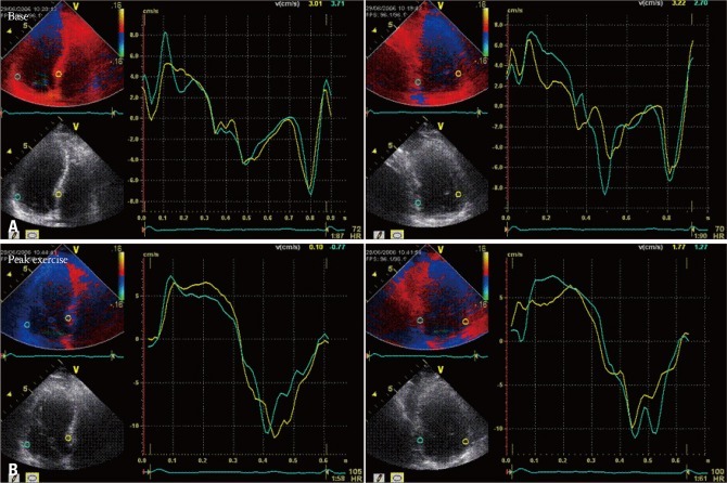
A total of 85 patients with hypertension who having exertional dyspnea and 30 control individuals were enrolled. Exercise stress echocardiography was performed using a symptom limited, multistage supine bicycle test. To evaluate the dyssynchrony of left ventricular (LV), we calculated the standard deviation (SD) of the averaged time-to-peak systolic velocity (TPs-SD, ms) of 12 middle and basal LV segments obtained from the three standard apical views at rest and peak exercise.
There was no significant difference in systolic blood pressure (BP) and heart rate between the two groups. TPs-SD was significantly higher in patients with LVH at rest (31.5 ± 12.1 vs. 22.0 ± 12.6 ms, p = 0.002) with exaggeration of the degree at peak exercise (39.0 ± 11.9 vs. 24.6 ± 13.3 ms, p < 0.001). Multiple regression analysis showed LV mass index was independently associated with LV dyssynchrony at peak exercise (β = 0.515, p = 0.001) when controlled for age, sex, and systolic BP at peak exercise.
Intraventricular systolic dyssynchrony during exercise is significantly associated with the degree of LVH in hypertensive patients.
高血压患者运动耐量受损并伴有呼吸困难很常见,这可能是由于运动期间心室非均匀激活加剧所致。因此,我们想要评估左心室肥厚(LVH)对运动期间收缩期心室内不同步的影响。
共纳入85例有劳力性呼吸困难的高血压患者和30例对照个体。采用症状限制的多级仰卧位自行车试验进行运动负荷超声心动图检查。为了评估左心室(LV)的不同步性,我们计算了静息和运动峰值时从三个标准心尖视图获得的12个左心室中间和基底节段的平均收缩期峰值速度时间(TPs-SD,毫秒)的标准差(SD)。
两组之间收缩压(BP)和心率无显著差异。LVH患者静息时TPs-SD显著更高(31.5±12.1对22.0±12.6毫秒,p = 0.002),运动峰值时程度加剧(39.0±11.9对24.6±13.3毫秒,p < 0.001)。多因素回归分析显示,在控制运动峰值时的年龄、性别和收缩压后,左心室质量指数与运动峰值时的左心室不同步独立相关(β = 0.515,p = 0.001)。
运动期间心室内收缩期不同步与高血压患者的LVH程度显著相关。