Department of Cell Physiology and Molecular Biophysics, Texas Tech University Health Sciences Center, Lubbock, TX 79430, USA.
J Gen Physiol. 2013 Feb;141(2):165-78. doi: 10.1085/jgp.201210876.
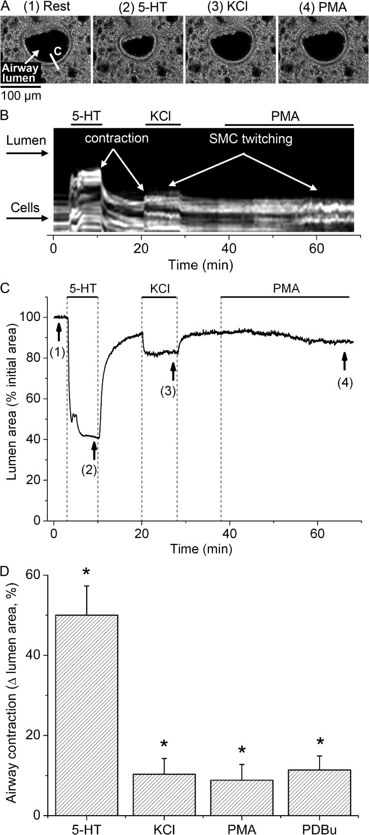
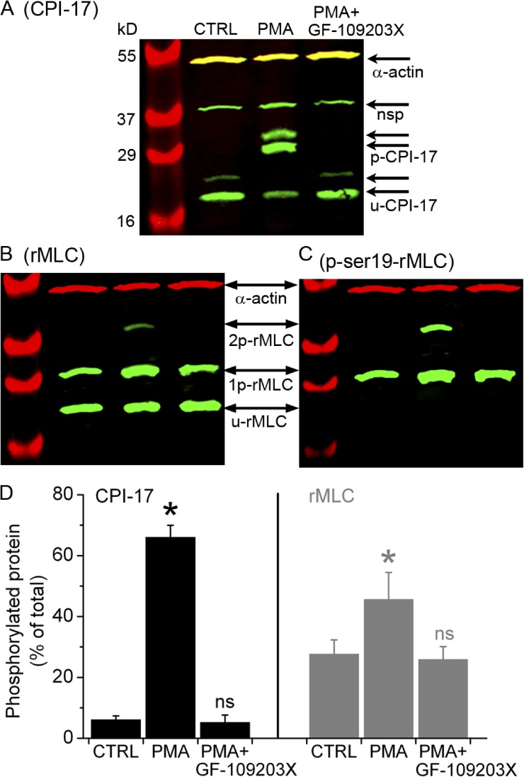
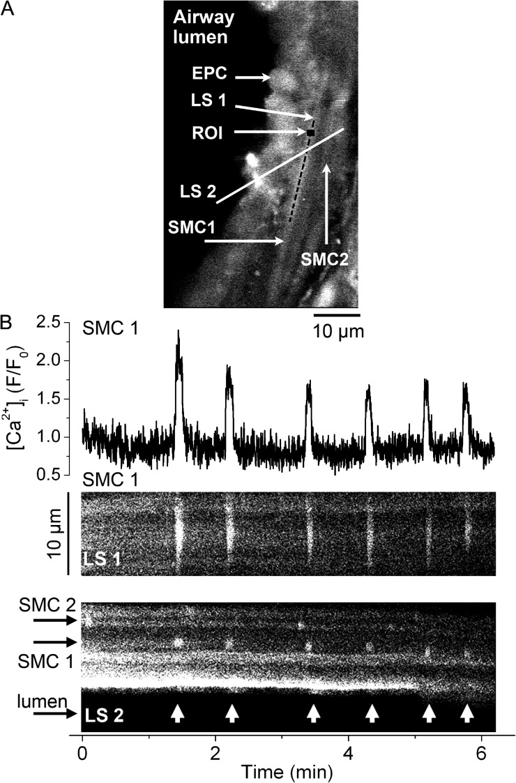
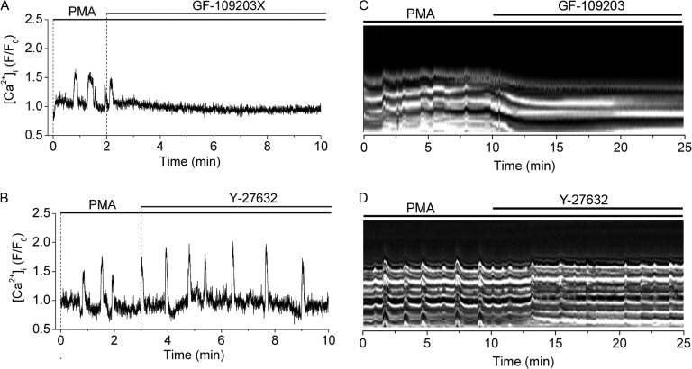
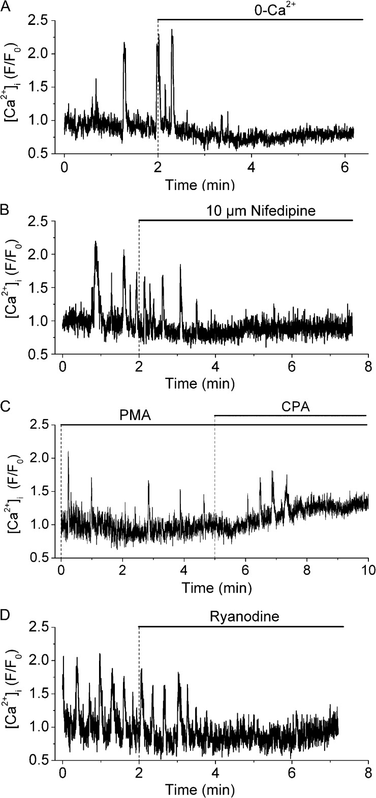
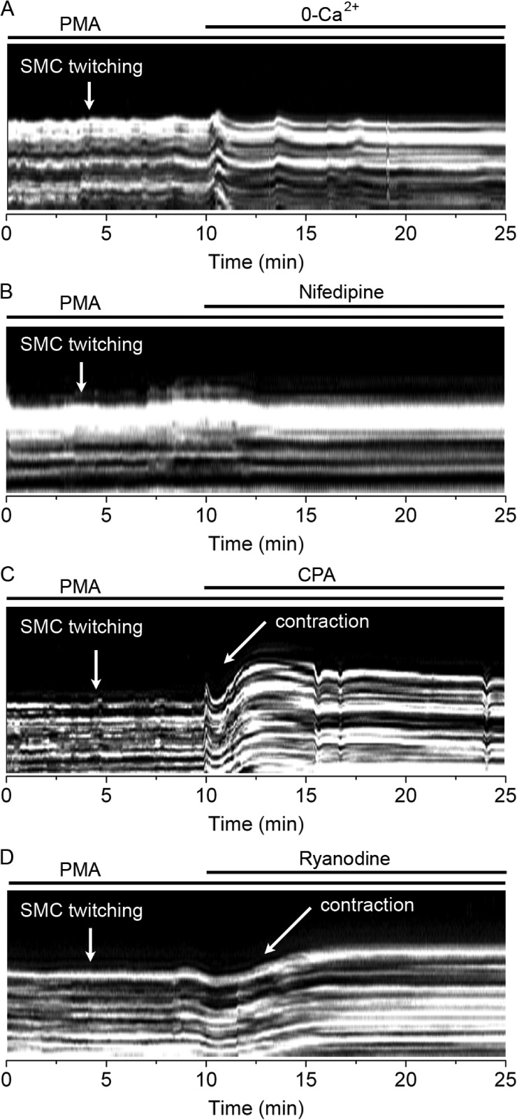
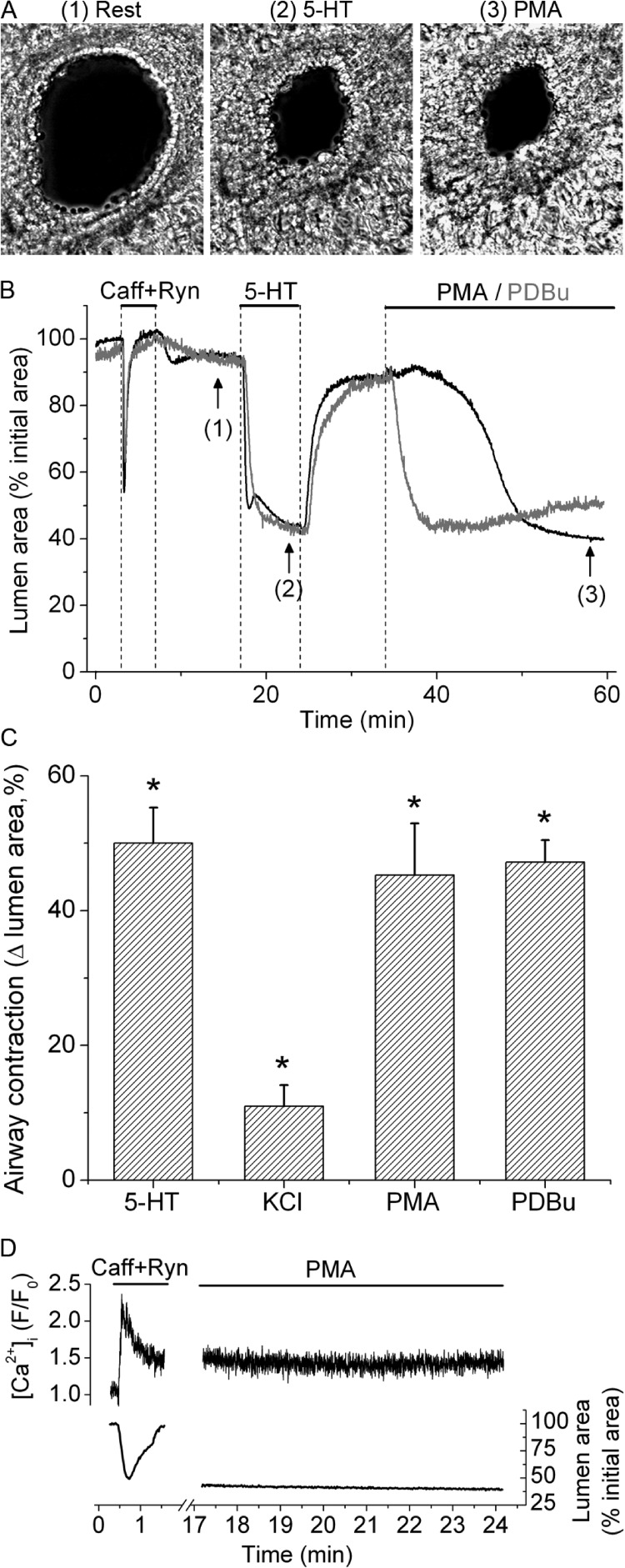
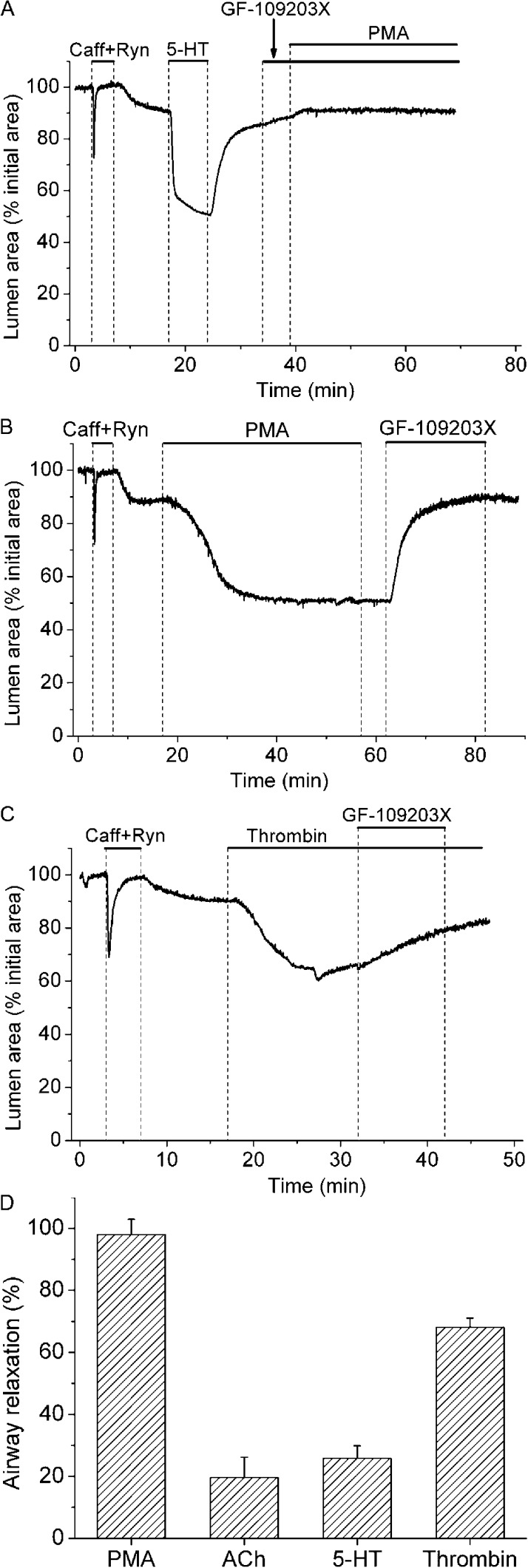
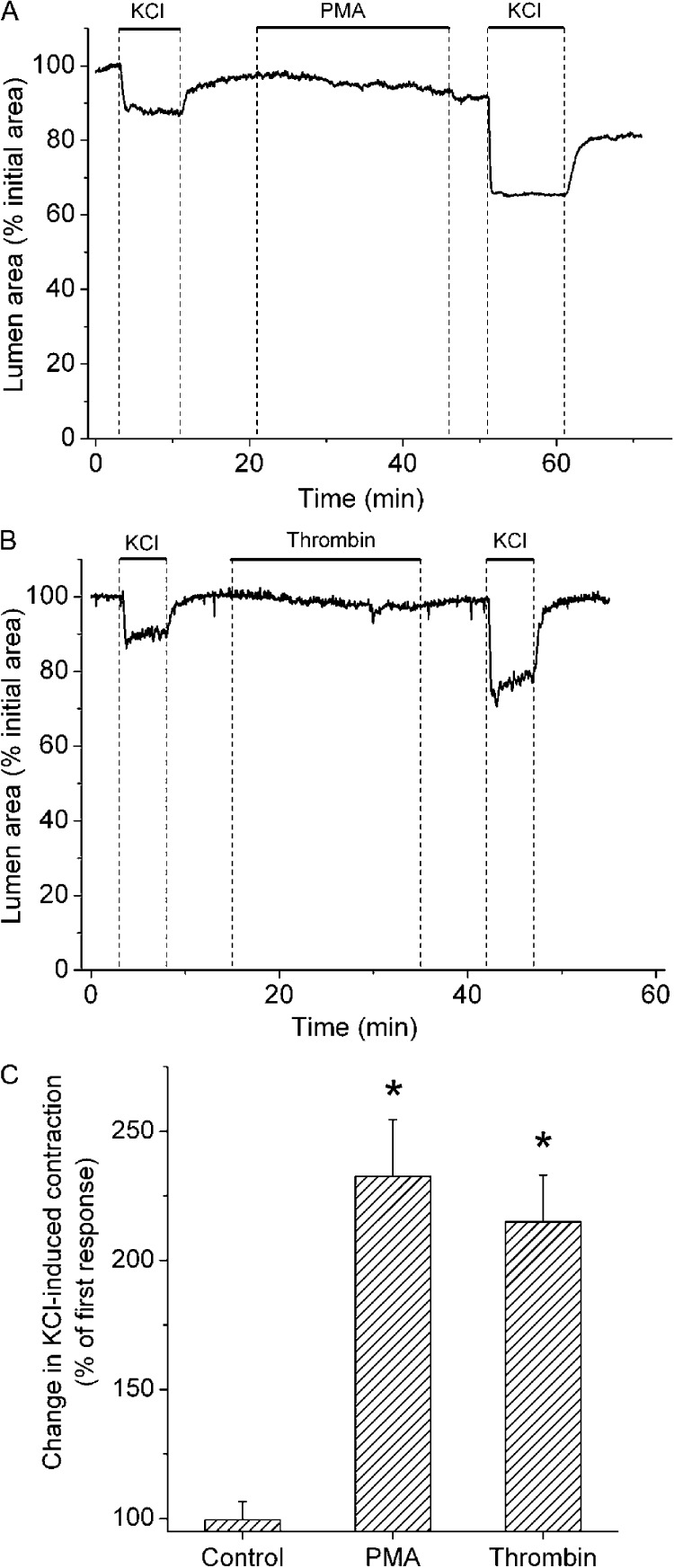
Protein kinase C (PKC) has been implicated in the regulation of smooth muscle cell (SMC) contraction and may contribute to airway hyperresponsiveness. Here, we combined optical and biochemical analyses of mouse lung slices to determine the effects of PKC activation on Ca(2+) signaling, Ca(2+) sensitivity, protein phosphorylation, and contraction in SMCs of small intrapulmonary airways. We found that 10 µM phorbol-12-myristate-13-acetate or 1 µM phorbol 12,13-dibutyrate induced repetitive, unsynchronized, and transient contractions of the SMCs lining the airway lumen. These contractions were associated with low frequency Ca(2+) oscillations in airway SMCs that resulted from Ca(2+) influx through L-type voltage-gated Ca(2+) channels and the subsequent release of Ca(2+) from intracellular stores through ryanodine receptors. Phorbol ester stimulation of lung slices in which SMC intracellular Ca(2+) concentration (Ca(2+)) was "clamped" at a high concentration induced strong airway contraction, indicating that PKC mediated sensitization of the contractile response to Ca(2+). This Ca(2+) sensitization was accompanied by phosphorylation of both the PKC-potentiated PP1 inhibitory protein of 17 kD (CPI-17) and the regulatory myosin light chain. Thrombin, like the phorbol esters, induced a strong Ca(2+) sensitization that was inhibited by the PKC inhibitor GF-109203X and also potentiated airway contraction to membrane depolarization with KCl. In conclusion, we suggest that PKC activation in small airways leads to both the generation of Ca(2+) oscillations and strong Ca(2+) sensitization; agents associated with airway inflammation, such as thrombin, may activate this pathway to sensitize airway smooth muscle to agonists that cause membrane depolarization and Ca(2+) entry and induce airway hyperresponsiveness.
蛋白激酶 C(PKC)已被牵涉到调节平滑肌细胞(SMC)收缩,并可能导致气道高反应性。在这里,我们结合光学和生化分析的小鼠肺切片,以确定 PKC 激活对 Ca(2 + )信号,Ca(2 + )敏感性,蛋白磷酸化和收缩的影响,在小的肺内气道的平滑肌细胞。我们发现,10 μM 佛波醇-12-肉豆蔻酸-13-醋酸酯或 1 μM 佛波醇 12,13-二丁酸诱导重复性,非同步和短暂的气道平滑肌细胞的收缩。这些收缩与低频率 Ca(2 + )振荡相关联,在气道平滑肌细胞中的 Ca(2 + )内流通过 L 型电压门控 Ca(2 + )通道和随后通过肌醇 1,4,5-三磷酸受体从细胞内储存中释放 Ca(2 + )引起。肺切片中 PKC 刺激,其中平滑肌细胞内 Ca(2 + )浓度([Ca(2 + )](i))被“夹”在高浓度诱导强气道收缩,表明 PKC 介导的收缩反应的敏感性增加对[Ca(2 + )](i)。这种 Ca(2 + )敏感性增加伴随着 PKC 增强的 17kD 蛋白磷酸酶 1 抑制蛋白(CPI-17)和调节肌球蛋白轻链的磷酸化。凝血酶,像佛波醇酯一样,诱导强烈的 Ca(2 + )敏感性增加,该增加被 PKC 抑制剂 GF-109203X 抑制,并且还增强了对 KCl 引起的膜去极化的气道收缩作用。总之,我们认为,小气道中的 PKC 激活导致 Ca(2 + )振荡的产生和强烈的 Ca(2 + )敏感性增加;与气道炎症相关的因子,如凝血酶,可能激活该途径,使气道平滑肌对引起膜去极化和 Ca(2 + )内流并诱导气道高反应性的激动剂敏感。