Department of Tumor Immunology, Radboud University Nijmegen Medical Centre, 6500 HB Nijmegen, The Netherlands.
Nat Commun. 2013;4:1412. doi: 10.1038/ncomms2402.
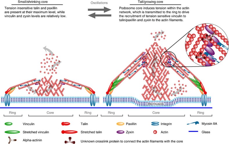
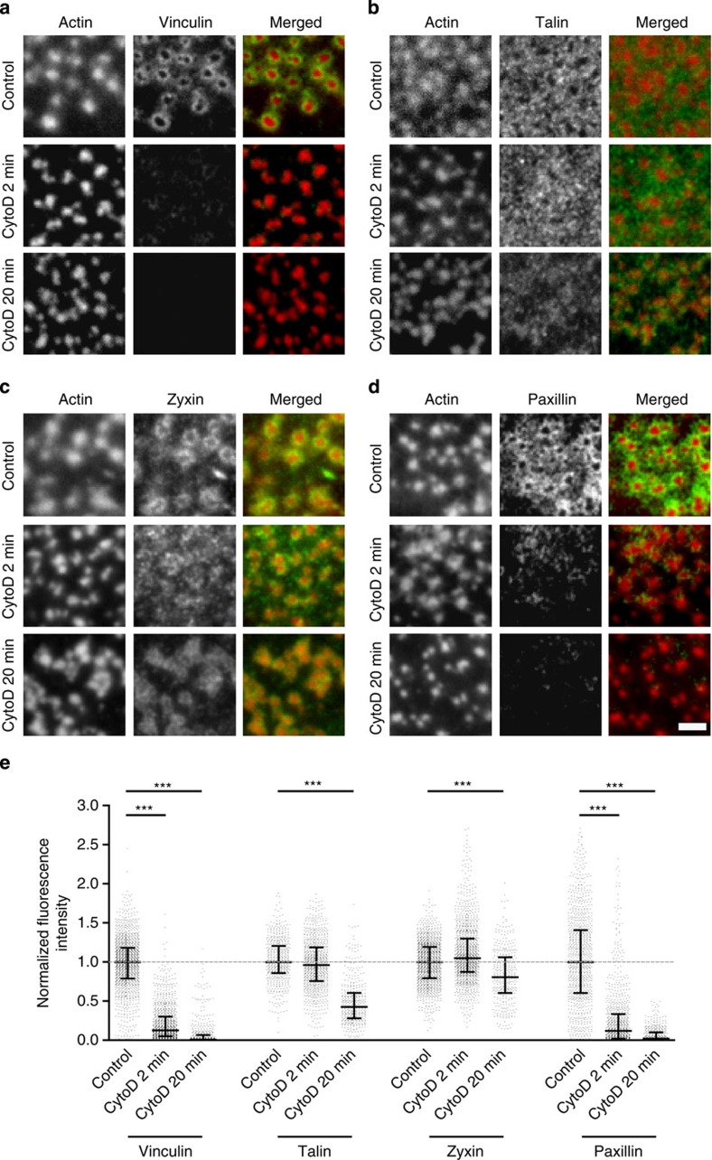
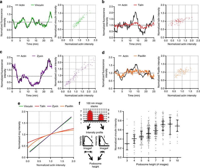
Tissue-resident dendritic cells patrol for foreign antigens while undergoing slow mesenchymal migration. Using actomyosin-based structures called podosomes, dendritic cells probe and remodel extracellular matrix topographical cues. Podosomes comprise an actin-rich protrusive core surrounded by an adhesive ring of integrins, cytoskeletal adaptor proteins and actin network filaments. Here we reveal how the integrity and dynamics of protrusive cores and adhesive rings are coordinated by the actomyosin apparatus. Core growth by actin polymerization induces podosome protrusion and provides tension within the actin network filaments. The tension transmitted to the ring recruits vinculin and zyxin and preserves overall podosome integrity. Conversely, myosin IIA contracts the actin network filaments and applies tension to the vinculin molecules bound, counterbalancing core growth and eventually reducing podosome size and protrusion. We demonstrate a previously unrecognized interplay between actin and myosin IIA in podosomes, providing novel mechanistic insights into how actomyosin-based structures allow dendritic cells to sense the extracellular environment.
组织驻留树突状细胞在进行缓慢的间充质迁移时巡逻寻找外来抗原。树突状细胞利用称为足突的肌动蛋白为基础的结构来探测和重塑细胞外基质的地形线索。足突由富含肌动蛋白的突起核心组成,周围是整合素、细胞骨架衔接蛋白和肌动蛋白网络丝的黏附环。在这里,我们揭示了肌动球蛋白装置如何协调突起核心和黏附环的完整性和动态性。肌动蛋白聚合诱导的核心生长引起足突突起,并在肌动蛋白网络丝内产生张力。传递到环上的张力募集 vinculin 和 zyxin,并保持足突的整体完整性。相反,肌球蛋白 IIA 收缩肌动蛋白网络丝,并对结合的 vinculin 分子施加张力,从而平衡核心生长,最终减小足突的大小和突起。我们证明了足突中肌动蛋白和肌球蛋白 IIA 之间以前未被认识到的相互作用,为肌动球蛋白基结构如何使树突状细胞能够感知细胞外环境提供了新的机制见解。