Department of Physiology II, Nara Medical University, School of Medicine, Kashihara, Nara, Japan.
PLoS One. 2013;8(1):e54814. doi: 10.1371/journal.pone.0054814. Epub 2013 Jan 31.
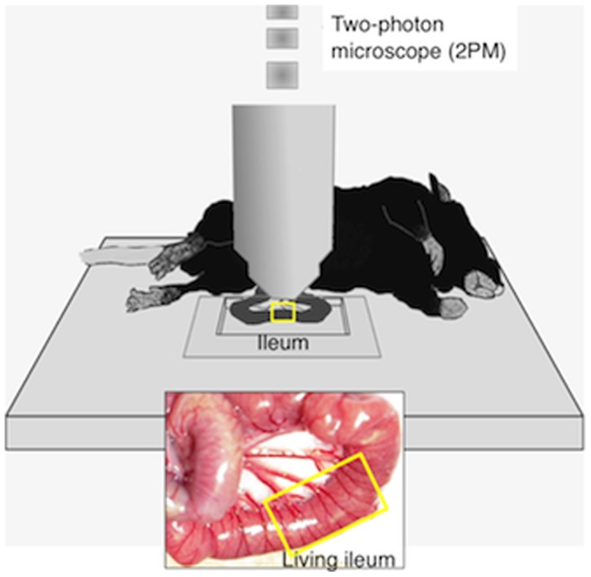
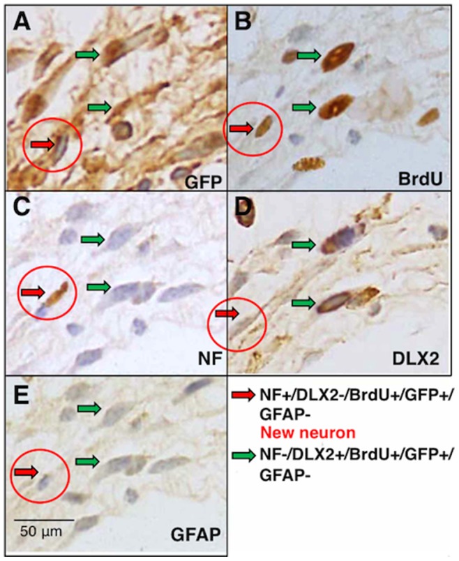
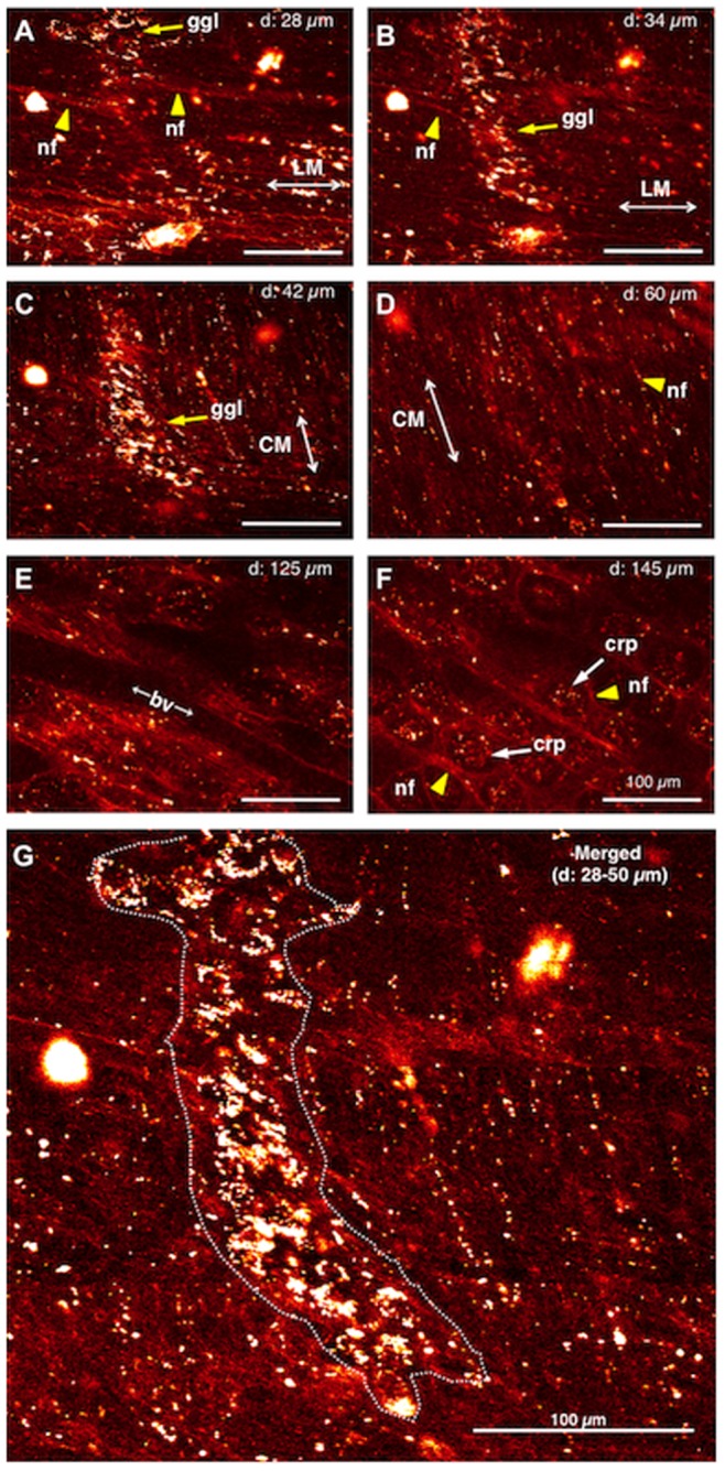
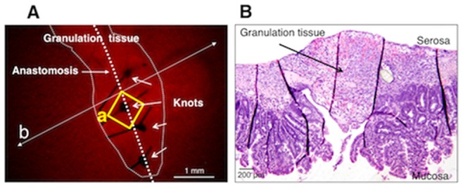
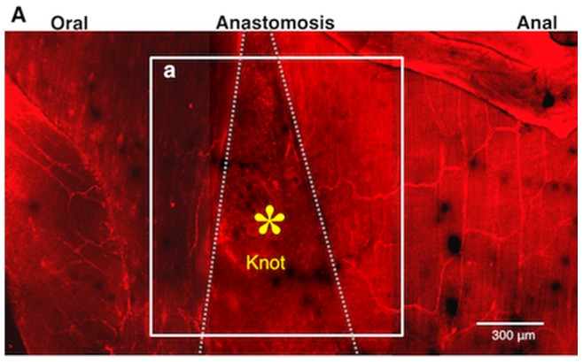
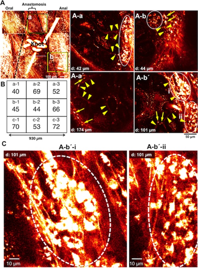
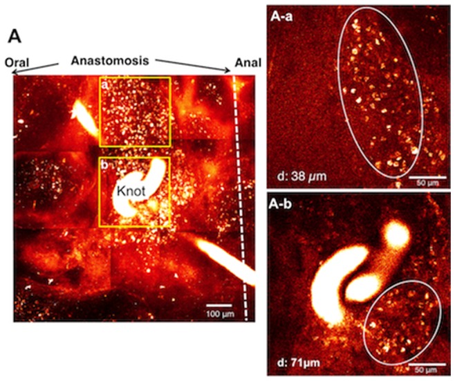
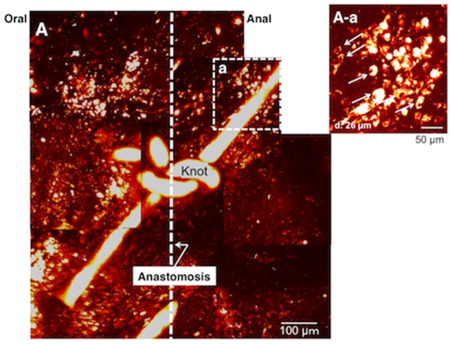
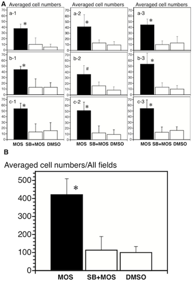
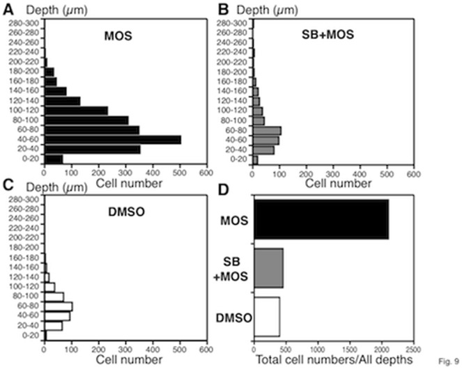
One of the challenges of using imaging techniques as a tool to study cellular physiology has been the inability to resolve structures that are not located near the surface of the preparation. Nonlinear optical microscopy, in particular two photon-excited fluorescence microscopy (2PM), has overcome this limitation, providing deeper optical penetration (several hundred µm) in ex vivo and in vivo preparations. We have used this approach in the gut to achieve the first in vivo imaging of enteric neurons and nerve fibers in the mucosa, submucosa, submucosal and myenteric plexuses, and circular and longitudinal muscles of the small intestine in H-line: Thy1 promoter GFP mice. Moreover, we obtained clear three-dimensional imaging of enteric neurons that were newly generated after gut transection and reanastomosis. Neurogenesis was promoted by oral application of the 5-HT(4)-receptor agonist, mosapride citrate (MOS). The number of newly generated neurons observed in mice treated with MOS for one week was 421±89 per 864,900 µm(2) (n = 5), which was significantly greater than that observed in preparations treated with MOS plus an antagonist (113±76 per 864,900 µm(2)) or in 4 week vehicle controls (100±34 per 864,900 µm(2)) (n = 4 both). Most neurons were located within 100 µm of the surface. These results confirm that activation of enteric neural 5-HT(4)-receptor by MOS promotes formation of new enteric neurons. We conclude that in vivo 2PM imaging made it possible to perform high-resolution deep imaging of the living mouse whole gut and reveal formation of new enteric neurons promoted by 5-HT(4)-receptor activation.
使用成像技术作为研究细胞生理学的工具之一的挑战之一是无法解析不在制备物表面附近的结构。非线性光学显微镜,特别是双光子激发荧光显微镜(2PM),克服了这一限制,在离体和体内制备物中提供了更深的光学穿透深度(数百 µm)。我们已经在肠道中使用这种方法来实现第一个活体成像,即在 H 线:Thy1 启动子 GFP 小鼠的粘膜、黏膜下层、黏膜下层和肌间神经丛以及小肠的环形和纵向肌肉中成像肠神经元和神经纤维。此外,我们获得了在肠道横断和再吻合后新生成的肠神经元的清晰三维成像。通过口服应用 5-HT(4)-受体激动剂莫沙必利枸橼酸盐(MOS)促进了神经发生。用 MOS 治疗一周的小鼠中观察到的新生成神经元的数量为每 864900 µm² 421±89 个(n = 5),明显多于用 MOS 加拮抗剂(每 864900 µm² 113±76 个)处理的制剂(n = 5) 4 周载体对照(每 864900 µm² 100±34 个)(n = 4)。大多数神经元位于表面 100 µm 以内。这些结果证实,MOS 激活肠神经元 5-HT(4)-受体可促进新肠神经元的形成。我们得出结论,体内 2PM 成像使对活体小鼠整个肠道进行高分辨率深度成像成为可能,并揭示了 5-HT(4)-受体激活促进新肠神经元形成。