Department of Genetics, Hyogo College of Medicine, 1-1 Mukogawa-cho, Nishinomiya, Hyogo 663-8501, Japan.
Int J Mol Sci. 2013 Feb 5;14(2):3215-27. doi: 10.3390/ijms14023215.
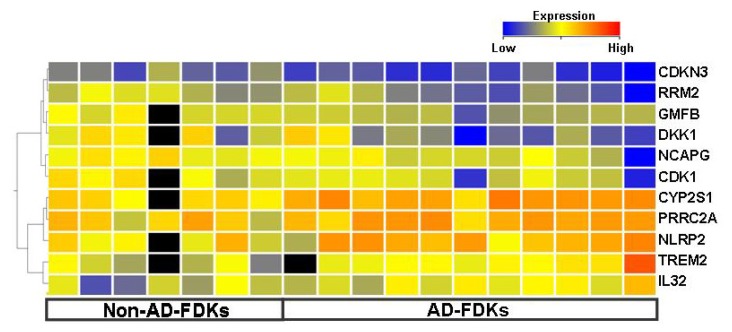
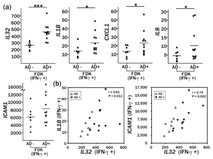
We cultured human hair follicle-derived keratinocytes (FDKs) from plucked hairs. To gain insight into gene expression signatures that can distinguish atopic dermatitis from non-atopic controls without skin biopsies, we undertook a comparative study of gene expression in FDKs from adult donors with atopic dermatitis and non-atopic donors. FDK primary cultures (atopic dermatitis, n = 11; non-atopic controls, n = 7) before and after interferon gamma (IFN-γ) treatment were used for microarray analysis and quantitative RT-PCR. Comparison of FDKs from atopic and non-atopic donors indicated that the former showed activated pathways with innate immunity and decreased pathways of cell growth, as indicated by increased NLRP2 expression and decreased DKK1 expression, respectively. Treatment with IFN-γ induced the enhanced expression of IL32, IL1B, IL8, and CXCL1 in the cells from atopic donors compared to that in cells from non-atopic donors at 24 h after treatment. IL1B expression in FDKs after IFN-γ treatment correlated with IL32 expression. We hypothesized that overexpression of IL32 in hair follicle keratinocytes of patients with atopic dermatitis would lead to the excessive production of pro-IL1β and that the activation of IL1β from pro-IL1β by inflammasome complex, in which NLRP2 protein might be involved, would be augmented. This is the first report to show enhanced induction of cytokine/chemokine genes by IFN-γ in atopic dermatitis using cultured FDKs.
我们从拔出的毛发中培养了人毛囊来源的角质形成细胞(FDK)。为了深入了解无需皮肤活检即可区分特应性皮炎和非特应性皮炎对照的基因表达特征,我们对特应性皮炎和非特应性皮炎供体的 FDK 进行了比较基因表达研究。使用干扰素γ(IFN-γ)处理前后的 FDK 原代培养物(特应性皮炎,n=11;非特应性皮炎对照,n=7)进行了微阵列分析和定量 RT-PCR。特应性皮炎和非特应性皮炎供体的 FDK 比较表明,前者显示出固有免疫激活途径和细胞生长途径减少,分别表现为 NLRP2 表达增加和 DKK1 表达减少。与非特应性皮炎供体的细胞相比,IFN-γ处理后,特应性皮炎供体的细胞中在 24 小时后诱导表达的细胞因子/趋化因子基因增加,包括 IL32、IL1B、IL8 和 CXCL1。IFN-γ处理后的 FDK 中 IL1B 的表达与 IL32 的表达相关。我们假设特应性皮炎患者的毛囊角质形成细胞中 IL32 的过度表达会导致前体 IL1β的过度产生,并且由 NLRP2 蛋白参与的炎性小体复合物激活前体 IL1β 生成的 IL1β 会增强。这是首次使用培养的 FDK 报告显示特应性皮炎中 IFN-γ 增强细胞因子/趋化因子基因的诱导。