Univ Paris Diderot, Sorbonne Paris Cité, Laboratory of Molecular and Cellular Responses to Xenobiotics, Unit of Functional and Adaptive Biology (BFA) EAC CNRS 4413, 5 rue Thomas Mann, Paris 75 013, France.
Part Fibre Toxicol. 2013 Feb 6;10:2. doi: 10.1186/1743-8977-10-2.
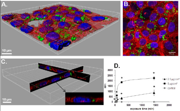
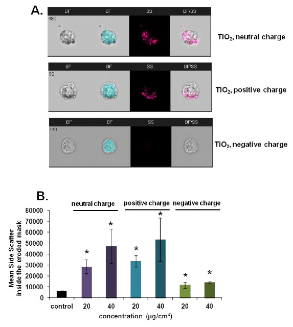
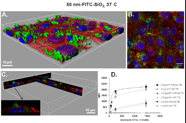
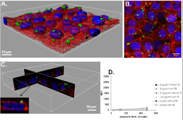
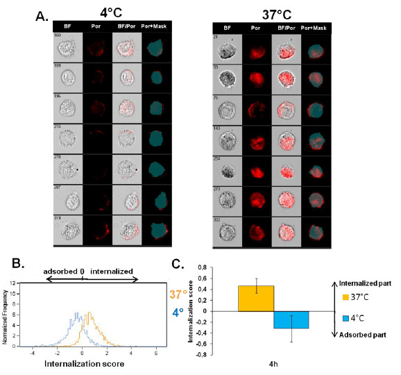
The uptake of nanoparticles (NPs) by cells remains to be better characterized in order to understand the mechanisms of potential NP toxicity as well as for a reliable risk assessment. Real NP uptake is still difficult to evaluate because of the adsorption of NPs on the cellular surface.
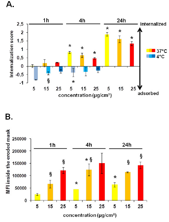
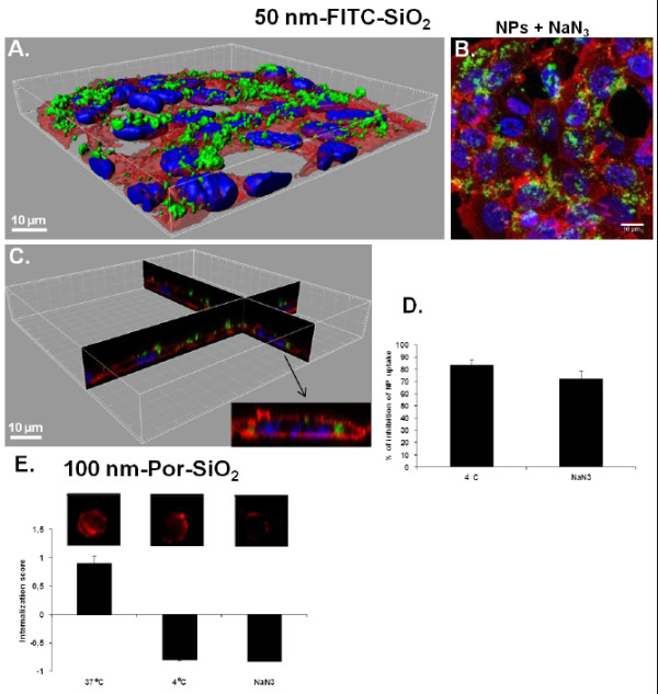
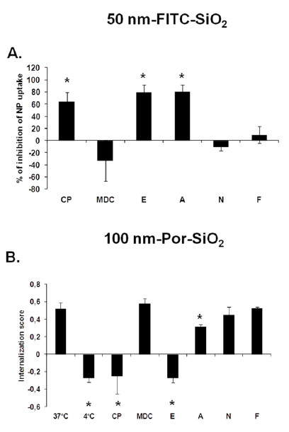
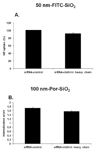
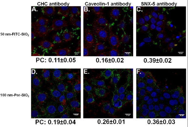
Here we used two approaches to distinguish adsorbed fluorescently labeled NPs from the internalized ones. The extracellular fluorescence was either quenched by Trypan Blue or the uptake was analyzed using imaging flow cytometry. We used this novel technique to define the inside of the cell to accurately study the uptake of fluorescently labeled (SiO2) and even non fluorescent but light diffracting NPs (TiO2). Time course, dose-dependence as well as the influence of surface charges on the uptake were shown in the pulmonary epithelial cell line NCI-H292. By setting up an integrative approach combining these flow cytometric analyses with confocal microscopy we deciphered the endocytic pathway involved in SiO2 NP uptake. Functional studies using energy depletion, pharmacological inhibitors, siRNA-clathrin heavy chain induced gene silencing and colocalization of NPs with proteins specific for different endocytic vesicles allowed us to determine macropinocytosis as the internalization pathway for SiO2 NPs in NCI-H292 cells.
The integrative approach we propose here using the innovative imaging flow cytometry combined with confocal microscopy could be used to identify the physico-chemical characteristics of NPs involved in their uptake in view to redesign safe NPs.
为了更好地了解潜在纳米颗粒毒性的作用机制以及进行可靠的风险评估,细胞对纳米颗粒(NPs)的摄取仍需进一步研究。由于 NPs 会吸附在细胞表面,因此很难评估真正的 NP 摄取量。
在这里,我们使用了两种方法来区分吸附在细胞表面的荧光标记 NPs 和内化的 NPs。通过锥虫蓝将细胞外荧光淬灭,或使用成像流式细胞术分析摄取情况。我们使用这项新技术来定义细胞内部,以准确研究荧光标记的(SiO2)和甚至是非荧光但光散射的 NPs(TiO2)的摄取情况。我们在肺上皮细胞系 NCI-H292 中研究了时间进程、剂量依赖性以及表面电荷对摄取的影响。通过建立一种整合方法,将这些流式细胞术分析与共聚焦显微镜相结合,我们阐明了涉及 SiO2 NP 摄取的内吞途径。使用能量耗竭、药理学抑制剂、siRNA-网格蛋白重链诱导基因沉默和 NPs 与不同内吞小泡的蛋白质共定位进行的功能研究,使我们能够确定巨胞饮作用是 NCI-H292 细胞内化 SiO2 NPs 的途径。
我们在这里提出的使用创新的成像流式细胞术结合共聚焦显微镜的综合方法,可用于确定与 NP 摄取相关的物理化学特性,以便重新设计安全的 NPs。