Institute of Molecular and Cell Biology, A-STAR, Singapore, Singapore.
PLoS One. 2013;8(2):e56219. doi: 10.1371/journal.pone.0056219. Epub 2013 Feb 7.
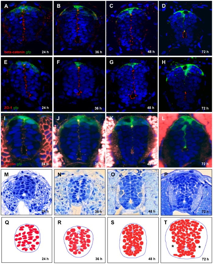
Neurulation is driven by apical constriction of actomyosin cytoskeleton resulting in conversion of the primitive lumen into the central canal in a mechanism driven by F-actin constriction, cell overcrowding and buildup of axonal tracts. The roof plate of the neural tube acts as the dorsal morphogenetic center and boundary preventing midline crossing by neural cells and axons.
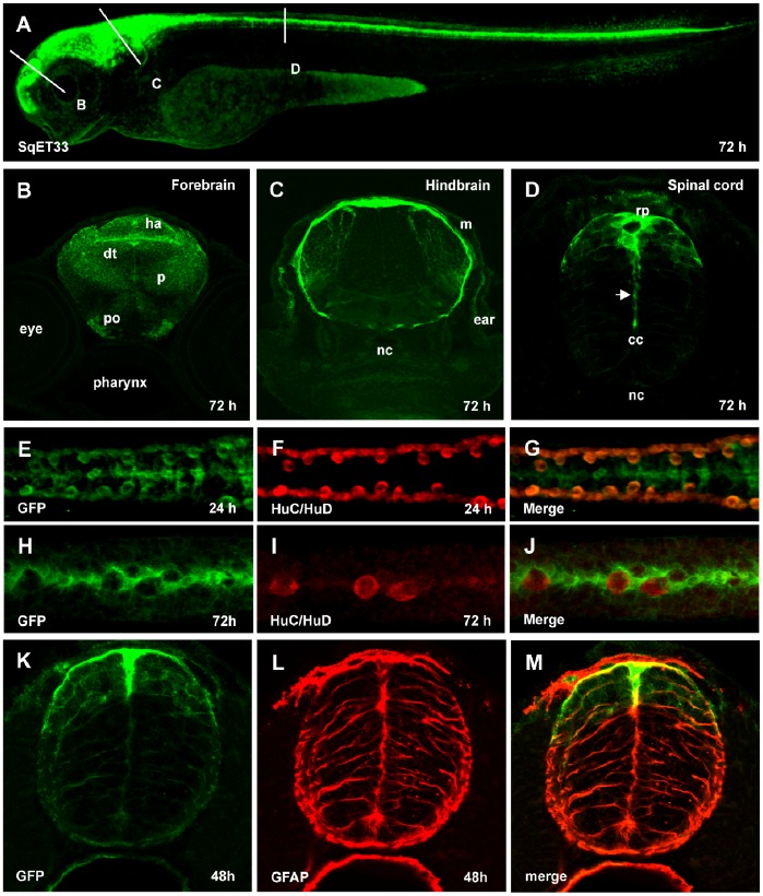
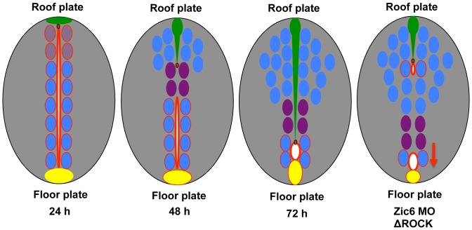
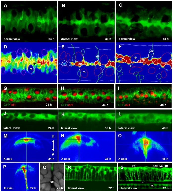
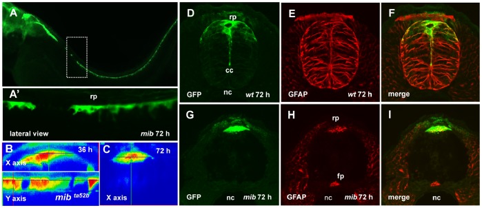
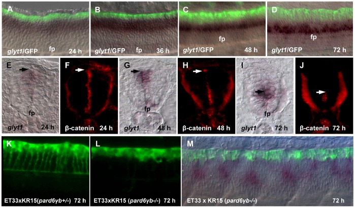
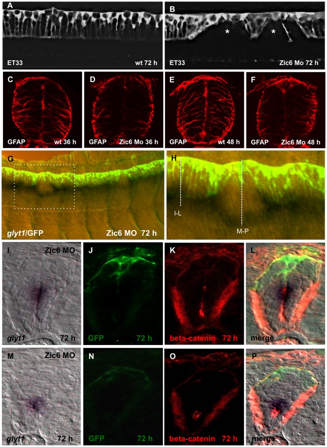
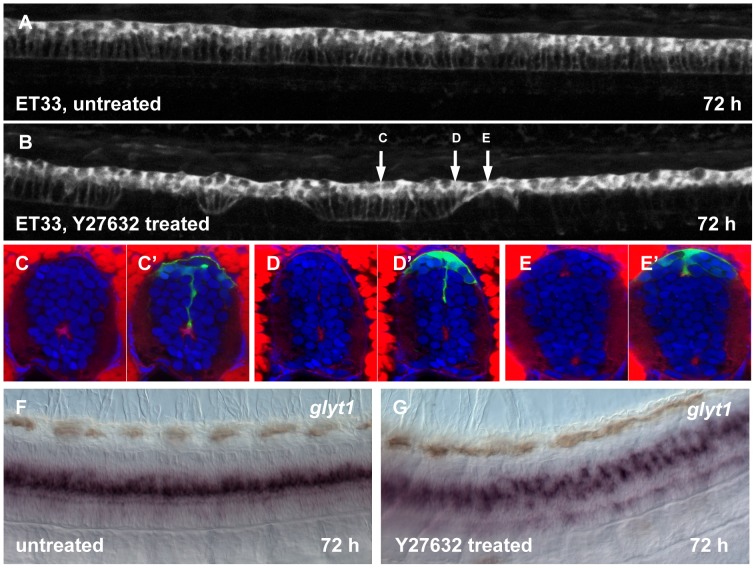
METHODOLOGY/PRINCIPAL FINDINGS: The roof plate zebrafish transgenics expressing cytosolic GFP were used to study and describe development of this structure in vivo for a first time ever. The conversion of the primitive lumen into the central canal causes significant morphogenetic changes of neuroepithelial cells in the dorsal neural tube. We demonstrated that the roof plate cells stretch along the D-V axis in parallel with conversion of the primitive lumen into central canal and its ventral displacement. Importantly, the stretching of the roof plate is well-coordinated along the whole spinal cord and the roof plate cells extend 3× in length to cover 2/3 of the neural tube diameter. This process involves the visco-elastic extension of the roof place cytoskeleton and depends on activity of Zic6 and the Rho-associated kinase (Rock). In contrast, stretching of the floor plate is much less extensive.
CONCLUSIONS/SIGNIFICANCE: The extension of the roof plate requires its attachment to the apical complex of proteins at the surface of the central canal, which depends on activity of Zic6 and Rock. The D-V extension of the roof plate may change a range and distribution of morphogens it produces. The resistance of the roof plate cytoskeleton attenuates ventral displacement of the central canal in illustration of the novel mechanical role of the roof plate during development of the body axis.
神经管的神经发生是由肌动球蛋白细胞骨架的顶端收缩驱动的,导致原始管腔转化为中央管,这一机制是由 F-肌动蛋白收缩、细胞过度拥挤和轴突束的积累驱动的。神经管的顶板作为背侧形态发生中心和边界,防止神经细胞和轴突从中线穿过。
方法/主要发现:本研究首次使用表达细胞质 GFP 的顶板斑马鱼转基因品系,在体内研究和描述了这一结构的发育。原始管腔转化为中央管导致背侧神经管神经上皮细胞发生显著的形态发生变化。我们证明,顶板细胞沿着 D-V 轴平行于原始管腔转化为中央管及其腹侧位移而伸展。重要的是,顶板的伸展在整个脊髓上都得到了很好的协调,顶板细胞伸展了 3 倍,覆盖了神经管直径的 2/3。这个过程涉及到顶板细胞骨架的粘弹性伸展,并且依赖于 Zic6 和 Rho 相关激酶(Rock)的活性。相比之下,底板的伸展范围要小得多。
结论/意义:顶板的伸展需要其与中央管表面的顶端复合物蛋白附着,这取决于 Zic6 和 Rock 的活性。顶板的 D-V 伸展可能会改变它产生的形态发生素的范围和分布。顶板细胞骨架的阻力减弱了中央管的腹侧位移,说明了顶板在身体轴发育过程中的新机械作用。