Institute of Molecular and Cell Biology, Agency for Science, Technology and Research, Singapore, Singapore.
Translational Medicine Centre, The First Affiliated Hospital, Sun Yat-sen University, Guangzhou, China.
Cell Tissue Res. 2021 Feb;383(2):835-852. doi: 10.1007/s00441-020-03270-1. Epub 2020 Sep 9.
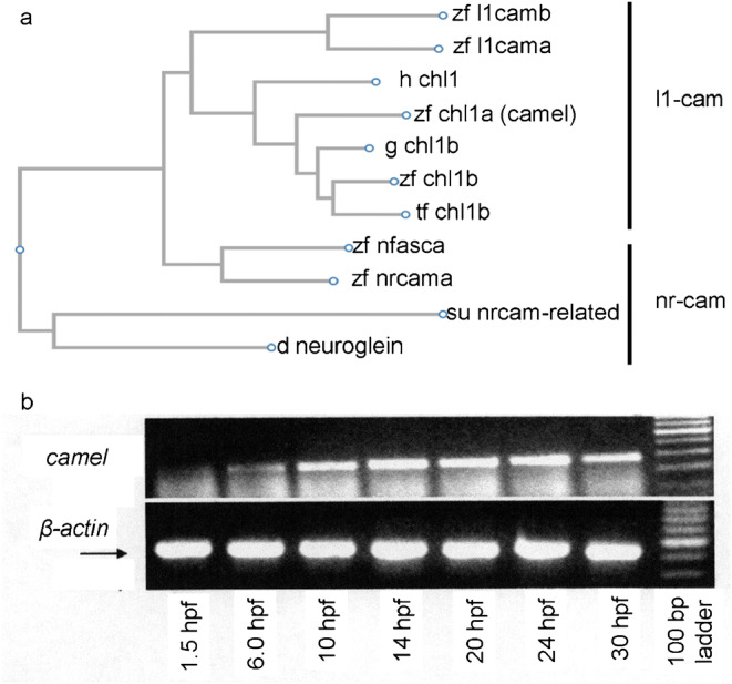
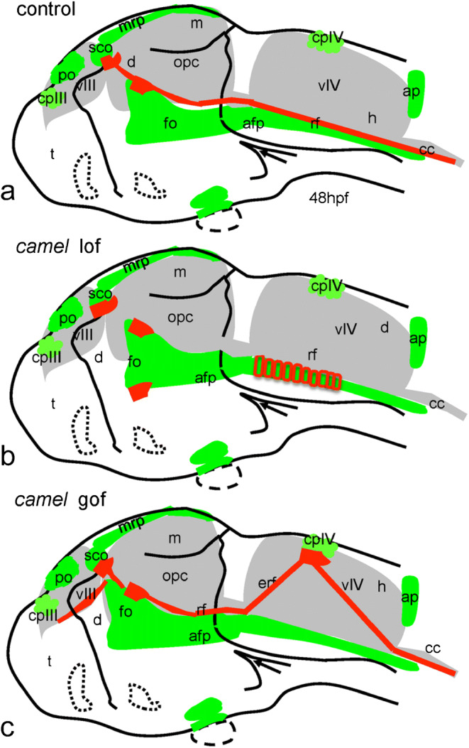
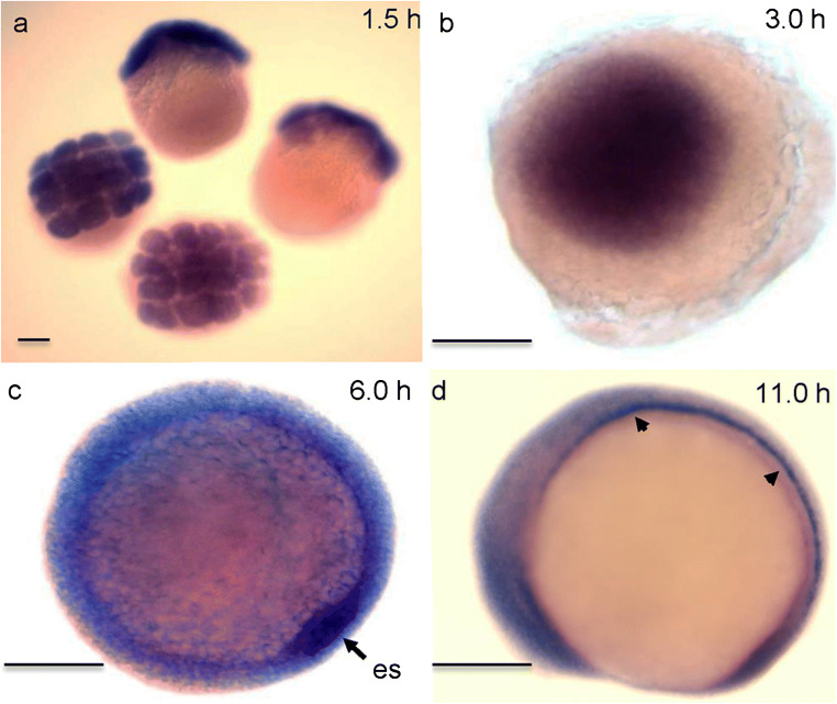
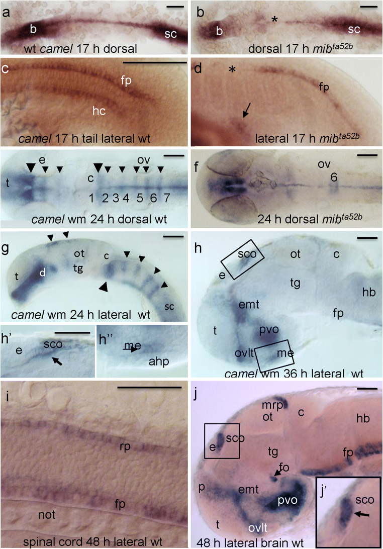
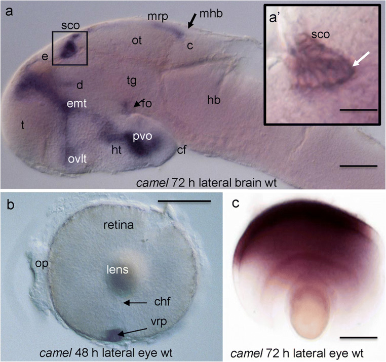
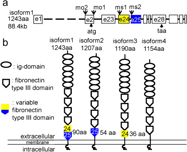
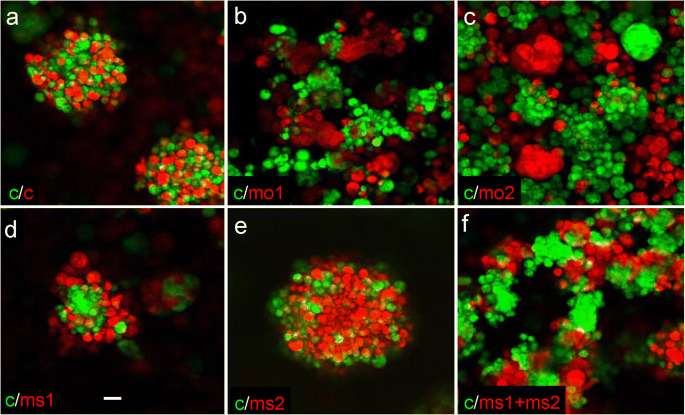
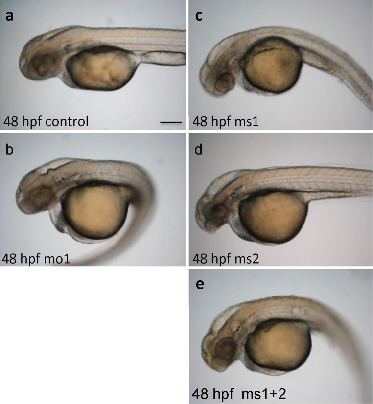
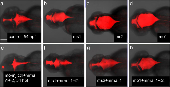
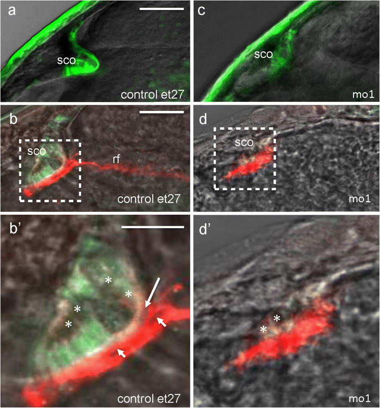
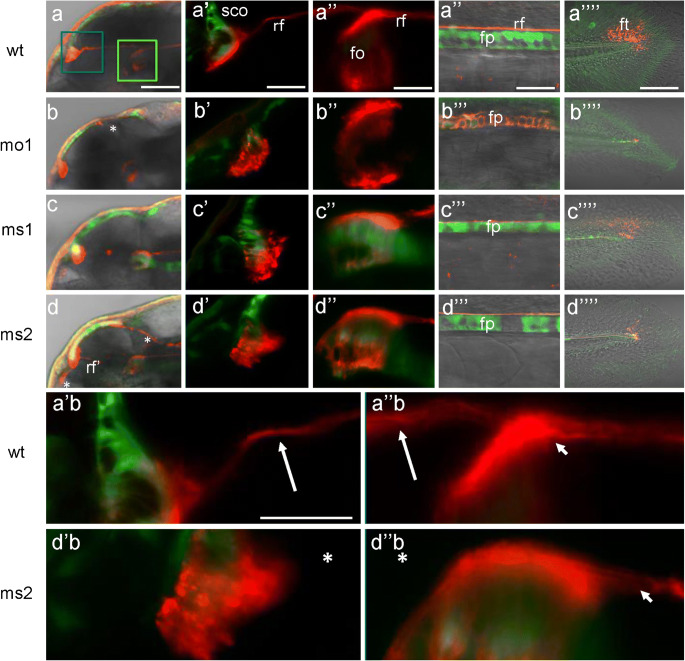
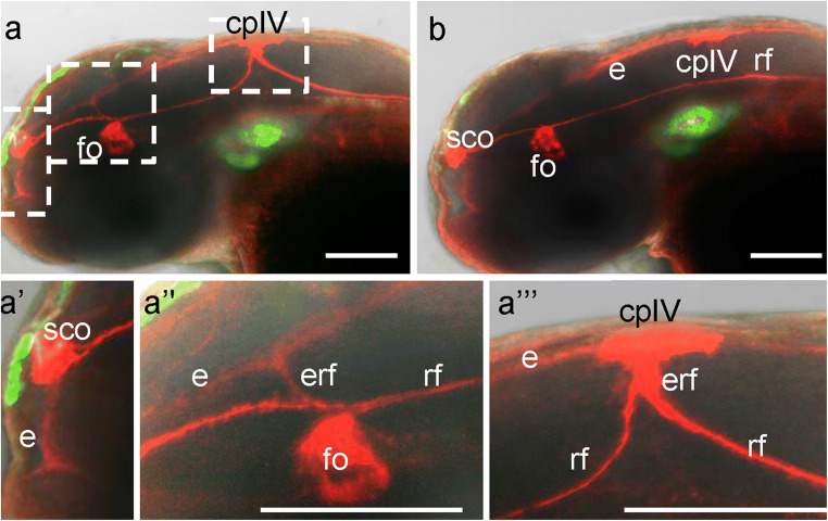
Development of the brain ventricular system of vertebrates and the molecular mechanisms involved are not fully understood. The developmental genes expressed in the elements of the brain ventricular system such as the ependyma and circumventricular organs act as molecular determinants of cell adhesion critical for the formation of brain ventricular system. They control brain development and function, including the flow of cerebrospinal fluid. Here, we describe the novel distantly related member of the zebrafish L1-CAM family of genes-camel. Whereas its maternal transcripts distributed uniformly, the zygotic transcripts demonstrate clearly defined expression patterns, in particular in the axial structures: floor plate, hypochord, and roof plate. camel expresses in several other cell lineages with access to the brain ventricular system, including the midbrain roof plate, subcommissural organ, organum vasculosum lamina terminalis, median eminence, paraventricular organ, flexural organ, and inter-rhombomeric boundaries. This expression pattern suggests a role of Camel in neural development. Several isoforms of Camel generated by differential splicing of exons encoding the sixth fibronectin type III domain enhance cell adhesion differentially. The antisense oligomer morpholino-mediated loss-of-function of Camel affects cell adhesion and causes hydrocephalus and scoliosis manifested via the tail curled down phenotype. The subcommissural organ's derivative-the Reissner fiber-participates in the flow of cerebrospinal fluid. The Reissner fiber fails to form upon morpholino-mediated Camel loss-of-function. The Camel mRNA-mediated gain-of-function causes the Reissner fiber misdirection. This study revealed a link between Chl1a/Camel and Reissner fiber formation, and this supports the idea that CHL1 is one of the scoliosis factors.
脊椎动物脑室系统的发育及其涉及的分子机制尚未完全阐明。在脑室系统的上皮和室周器官等元件中表达的发育基因作为细胞黏附的分子决定因素,对脑室系统的形成至关重要。它们控制着大脑的发育和功能,包括脑脊液的流动。在这里,我们描述了斑马鱼 L1-CAM 家族基因的一个新的远缘成员——骆驼。虽然其母本转录本均匀分布,但合子转录本表现出明确的表达模式,特别是在轴向结构中:基板、脊索和顶板。骆驼在其他几个进入脑室系统的细胞谱系中表达,包括中脑顶板、室管下器官、终板血管组织、正中隆起、室旁器官、弯曲器官和间脑节边界。这种表达模式表明 Camel 在神经发育中具有重要作用。由第六个纤维连接蛋白 III 结构域编码外显子差异剪接产生的几种 Camel 同工型差异增强细胞黏附。使用反义寡核苷酸介导的 Camel 功能丧失会影响细胞黏附,并导致通过尾巴卷曲向下表型表现出的脑积水和脊柱侧凸。室管下器官的衍生物——Reissner 纤维——参与脑脊液的流动。在反义寡核苷酸介导的 Camel 功能丧失时,Reissner 纤维无法形成。Camel mRNA 介导的功能获得导致 Reissner 纤维错位。这项研究揭示了 Chl1a/Camel 与 Reissner 纤维形成之间的联系,这支持了 CHL1 是脊柱侧凸因子之一的观点。