Laboratory of Medical Microbiology, Hellenic Pasteur Institute, Athens, Greece.
PLoS One. 2013;8(2):e56291. doi: 10.1371/journal.pone.0056291. Epub 2013 Feb 7.
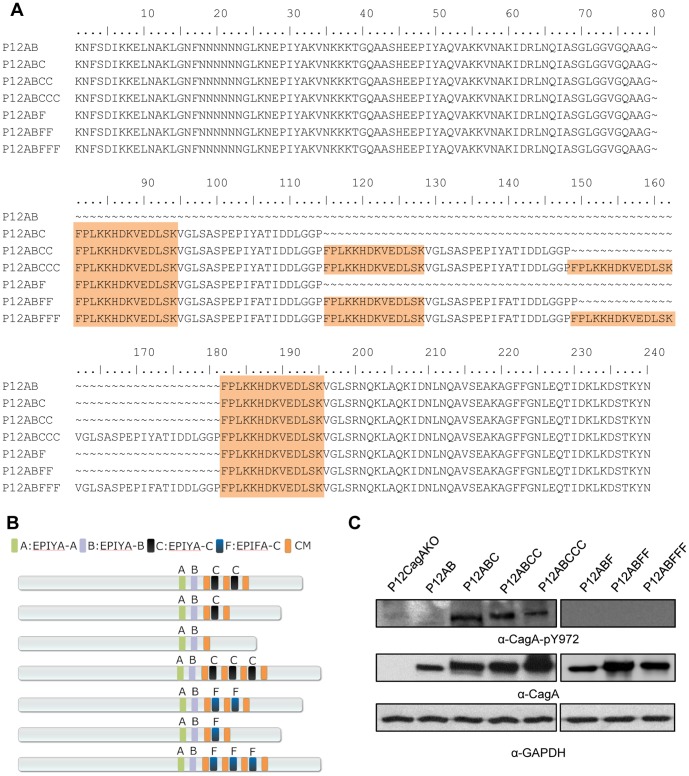
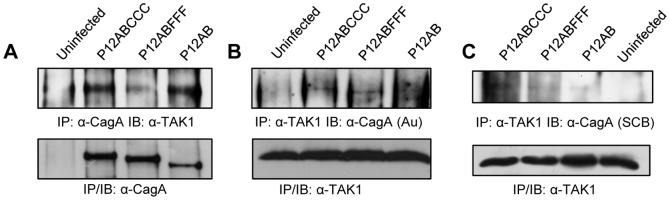
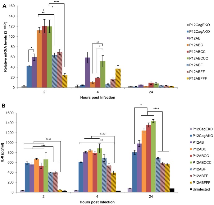
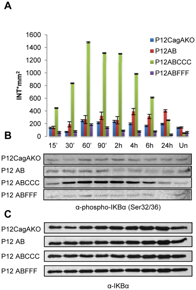
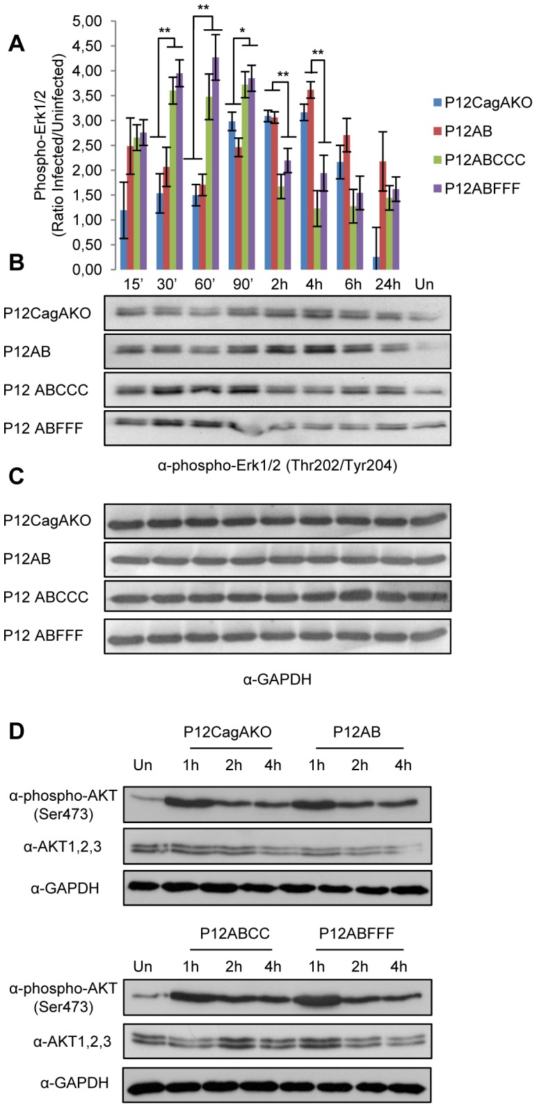
CagA protein contributes to pro-inflammatory responses during H. pylori infection, following its intracellular delivery to gastric epithelial cells. Here, we report for the first time in an isogenic background, on the subtle role of CagA phosphorylation on terminal EPIYA-C motifs in the transcriptional activation and expression of IL-8. We utilized isogenic H. pylori mutants of P12 reference strain, expressing CagA with varying number of EPIYA-C motifs and the corresponding phosphorylation defective EPIFA-C motifs while preserving intact the CM multimerization motifs. These mutants had been previously closely scrutinized in terms of type IV secretion system functionality, CagA translocation and its subsequent phosphorylation. Following infection of gastric epithelial cell lines, transcriptional activation of IL-8 gene and secreted IL-8 levels were found to be strictly dependent upon the functionality of the EPIYA-C phosphorylation motifs, as EPIFA-C phosphorylation-deficient CagA expression failed to induce full IL-8 transcriptional activity. Interestingly, levels of IL-8 gene activation and of secreted IL-8 were the same, irrespective of the number of EPIYA-C terminal repeats. We monitored IkBα phosphorylation and confirmed CagA involvement in NF-kB activation. Furthermore, we observed that presence of EPIYA-C functional phosphorylation motifs contributed to NF-kB activation. NF-kB upstream signaling events, such as early ERK1/2 and AKT activation were confirmed to be independent of EPIYA-C phosphorylation. On the contrary, use of TAK1 specific inhibitor 5Z-7-Oxozeaenol resulted in complete arrest of IL-8 secretion, in a dose-dependent manner, irrespective of CagA status. H. pylori-infected TAK1(-/-) mouse embryonic fibroblasts (MEFs) failed to induce NF-kB activity, unlike the respective control MEFs. CagA and TAK1 were found to immunoprecipitate together, irrespective of CagA EPIYA-C status, thus confirming earlier reports of TAK1 and CagA protein interaction. Our data suggest that CagA may potentially interfere with TAK1 activity during NF-kB activation for IL-8 induction in early H. pylori infection.
CagA 蛋白在 H. pylori 感染期间有助于引发促炎反应,这是通过其向胃上皮细胞内的递送来实现的。在这里,我们首次在同基因背景下报告了 CagA 在 EPIYA-C 末端基序上的磷酸化在 IL-8 的转录激活和表达中的微妙作用。我们利用 P12 参考菌株的同基因 H. pylori 突变体,表达具有不同数量的 EPIYA-C 基序和相应的磷酸化缺陷的 EPIFA-C 基序的 CagA,同时保持 CM 多聚化基序的完整。这些突变体在 IV 型分泌系统功能、CagA 易位及其随后的磷酸化方面已经进行了密切研究。在感染胃上皮细胞系后,发现 IL-8 基因的转录激活和分泌的 IL-8 水平严格依赖于 EPIYA-C 磷酸化基序的功能,因为 EPIFA-C 磷酸化缺陷的 CagA 表达未能诱导完全的 IL-8 转录活性。有趣的是,无论 EPIYA-C 末端重复的数量如何,IL-8 基因激活和分泌的 IL-8 水平都相同。我们监测了 IkBα 的磷酸化,并证实了 CagA 在 NF-kB 激活中的作用。此外,我们观察到 EPIYA-C 功能磷酸化基序的存在有助于 NF-kB 的激活。NF-kB 的上游信号事件,如早期的 ERK1/2 和 AKT 的激活,被证实与 EPIYA-C 磷酸化无关。相反,使用 TAK1 特异性抑制剂 5Z-7-Oxozeaenol 以剂量依赖性方式完全阻止了 IL-8 的分泌,而与 CagA 状态无关。与各自的对照 MEFs 不同,感染 H. pylori 的 TAK1(-/-) 鼠胚胎成纤维细胞 (MEFs) 未能诱导 NF-kB 活性。CagA 和 TAK1 被发现无论 CagA EPIYA-C 状态如何都能共同免疫沉淀,从而证实了之前关于 TAK1 和 CagA 蛋白相互作用的报道。我们的数据表明,在早期 H. pylori 感染期间,CagA 可能会干扰 NF-kB 激活过程中的 TAK1 活性,从而诱导 IL-8 的产生。