Department of Biomedical Sciences, NYIT College of Osteopathic Medicine, New York Institute of Technology, Old Westbury, NY 11568, USA.
J Transl Med. 2013 Feb 14;11:40. doi: 10.1186/1479-5876-11-40.
Left ventricular (LV) remodeling following large transmural myocardial infarction (MI) remains a pivotal clinical issue despite the advance of medical treatment over the past few decades. Identification of new medications to improve the remodeling process and prevent progression to heart failure after MI is critical. Thyroid hormones (THs) have been shown to improve LV function and remodeling in animals post-MI and in the human setting. However, changes in underlying cellular remodeling resulting from TH treatment are not clear.
MI was produced in adult female Sprague-Dawley rats by ligation of the left descending coronary artery. L-thyroxine (T4) pellet (3.3 mg, 60 days sustained release) was used to treat MI rats for 8 weeks. Isolated myocyte shape, arterioles, and collagen deposition in the non-infarcted area were measured at terminal study.
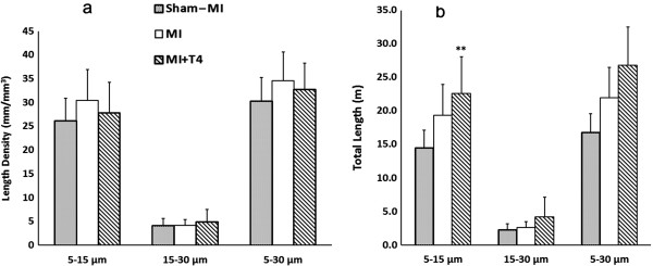
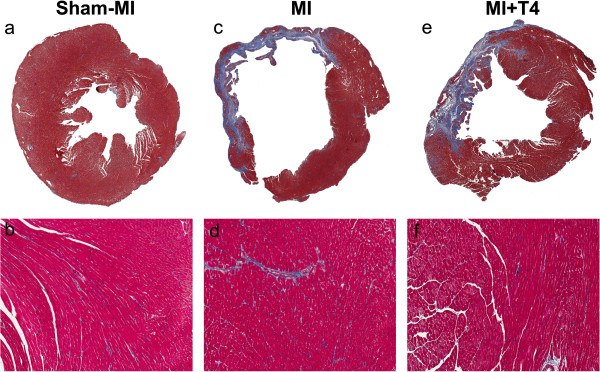
T4 treatment improved LV ±dp/dt, normalized TAU, and increased myocyte cross-sectional area without further increasing myocyte length in MI rats. T4 treatment increased the total LV tissue area by 34%, increased the non-infarcted tissue area by 41%, and increased the thickness of non-infarcted area by 36% in MI rats. However, myocyte volume accounted for only ~1/3 of the increase in myocyte mass in the non-infarct area, indicating the presence of more myocytes with treatment. T4 treatment tended to increase the total length of smaller arterioles (5 to 15 μm) proportional to LV weight increase and also decreased collagen deposition in the LV non-infarcted area. A tendency for increased metalloproteinase-2 (MMP-2) expression and tissue inhibitor of metalloproteinases (TIMPs) -1 to -4 expression was also observed in T4 treated MI rats.
These results suggest that long-term T4 treatment after MI has beneficial effects on myocyte, arteriolar, and collagen matrix remodeling in the non-infarcted area. Most importantly, results suggest improved survival of myocytes in the peri-infarct area.
尽管过去几十年医学治疗取得了进步,但左心室(LV)在大面积透壁性心肌梗死(MI)后的重塑仍然是一个关键的临床问题。寻找新的药物来改善重塑过程并预防 MI 后心力衰竭的进展至关重要。甲状腺激素(THs)已被证明可改善动物 MI 后和人类 MI 后的 LV 功能和重塑。然而,TH 治疗导致的潜在细胞重塑变化尚不清楚。
通过结扎左冠状动脉降支在成年雌性 Sprague-Dawley 大鼠中产生 MI。使用 L-甲状腺素(T4)丸(3.3mg,60 天持续释放)治疗 MI 大鼠 8 周。在终末研究中测量非梗死区的分离心肌细胞形状、小动脉和胶原沉积。
T4 治疗改善了 MI 大鼠的 LV ±dp/dt,使 TAU 正常化,并增加了心肌细胞的横截面积,而不进一步增加心肌细胞的长度。T4 治疗使 MI 大鼠的总 LV 组织面积增加了 34%,非梗死组织面积增加了 41%,非梗死区厚度增加了 36%。然而,心肌细胞体积仅占非梗死区心肌细胞质量增加的约 1/3,表明治疗后存在更多的心肌细胞。T4 治疗倾向于增加与 LV 重量增加成比例的较小(5 至 15μm)小动脉的总长度,同时减少 LV 非梗死区的胶原沉积。在 T4 治疗的 MI 大鼠中,还观察到金属蛋白酶-2(MMP-2)表达和组织抑制剂金属蛋白酶(TIMPs)-1 至 -4 表达的趋势增加。
这些结果表明,MI 后长期 T4 治疗对非梗死区心肌细胞、小动脉和胶原基质重塑具有有益作用。最重要的是,结果表明梗死区周围心肌细胞的存活率提高。