Department of Biochemistry, Biocenter, University of Wuerzburg, Wuerzburg, Germany.
PLoS One. 2013;8(2):e56317. doi: 10.1371/journal.pone.0056317. Epub 2013 Feb 18.
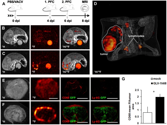
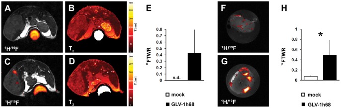
Oncolytic virotherapy of tumors is an up-coming, promising therapeutic modality of cancer therapy. Unfortunately, non-invasive techniques to evaluate the inflammatory host response to treatment are rare. Here, we evaluate (19)F magnetic resonance imaging (MRI) which enables the non-invasive visualization of inflammatory processes in pathological conditions by the use of perfluorocarbon nanoemulsions (PFC) for monitoring of oncolytic virotherapy.
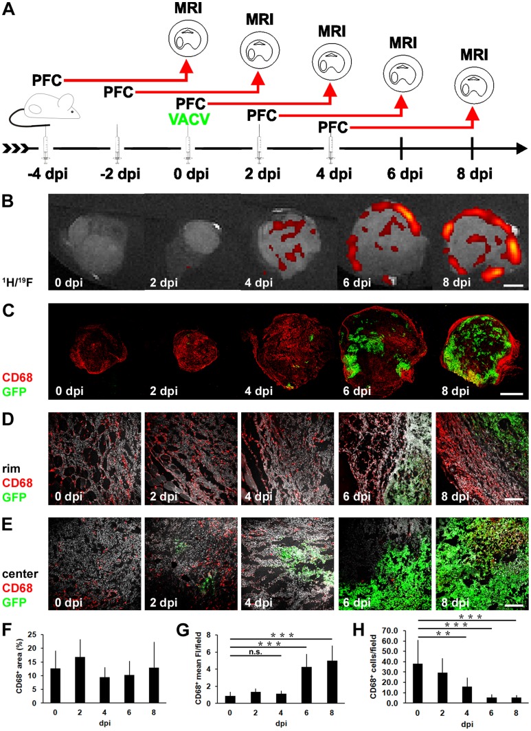
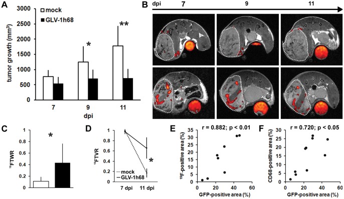
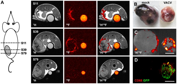
METHODOLOGY/PRINCIPAL FINDINGS: The Vaccinia virus strain GLV-1h68 was used as an oncolytic agent for the treatment of different tumor models. Systemic application of PFC emulsions followed by (1)H/(19)F MRI of mock-infected and GLV-1h68-infected tumor-bearing mice revealed a significant accumulation of the (19)F signal in the tumor rim of virus-treated mice. Histological examination of tumors confirmed a similar spatial distribution of the (19)F signal hot spots and CD68(+)-macrophages. Thereby, the CD68(+)-macrophages encapsulate the GFP-positive viral infection foci. In multiple tumor models, we specifically visualized early inflammatory cell recruitment in Vaccinia virus colonized tumors. Furthermore, we documented that the (19)F signal correlated with the extent of viral spreading within tumors.
CONCLUSIONS/SIGNIFICANCE: These results suggest (19)F MRI as a non-invasive methodology to document the tumor-associated host immune response as well as the extent of intratumoral viral replication. Thus, (19)F MRI represents a new platform to non-invasively investigate the role of the host immune response for therapeutic outcome of oncolytic virotherapy and individual patient response.
肿瘤溶瘤病毒治疗是一种有前途的癌症治疗方法。不幸的是,很少有非侵入性技术来评估治疗引起的炎症宿主反应。在这里,我们评估了(19)氟磁共振成像(MRI),通过使用全氟碳纳米乳液(PFC)可以非侵入性地可视化病理条件下的炎症过程,用于监测溶瘤病毒治疗。
方法/主要发现:使用牛痘病毒株 GLV-1h68 作为溶瘤剂治疗不同的肿瘤模型。对模拟感染和 GLV-1h68 感染的荷瘤小鼠进行 PFC 乳液系统应用后,(1)H/(19)F MRI 显示病毒治疗小鼠的肿瘤边缘有明显的(19)F 信号积聚。肿瘤的组织学检查证实了(19)F 信号热点和 CD68(+)-巨噬细胞的相似空间分布。因此,CD68(+)-巨噬细胞包裹 GFP 阳性病毒感染灶。在多种肿瘤模型中,我们特异性地可视化了痘苗病毒定植肿瘤中的早期炎症细胞募集。此外,我们证明(19)F 信号与肿瘤内病毒复制的程度相关。
结论/意义:这些结果表明(19)F MRI 是一种非侵入性方法,可以记录肿瘤相关的宿主免疫反应以及肿瘤内病毒复制的程度。因此,(19)F MRI 代表了一种新的平台,可以非侵入性地研究宿主免疫反应在溶瘤病毒治疗治疗效果和个体患者反应中的作用。