Defendenti Caterina, Atzeni Fabiola, Croce Anna Maria, Mussani Elena, Saibeni Simone, Bollani Simona, Grosso Silvia, Almasio Piero Luigi, Bruno Savino, Sarzi-Puttini Piercarlo
Laboratory Unit and Divisions of Fatebenefratelli Hospital, Milan, Italy.
BMC Clin Pathol. 2013 Mar 1;13:8. doi: 10.1186/1472-6890-13-8.
We have recently investigated the localisation of immunoglobulin-producing cells (IPCs) in inflamed intestinal tissue samples from patients with inflammatory bowel disease (IBD), and identified two main patterns of B lymphocyte infiltration: one characterised by the moderate strong stromal localisation of small B1 cell-like IgM+/CD79+/CD20-/CD21-/CD23-/CD5 ± IPCs, and the other by the peri-glandular localisation of IPCs with irregular nuclei that had surface markers specific for a B cell subset (IgM and CD79), but quantitative differences in their λ and κ chains. The same patients were also tested for CD15+ receptors, which were localised on inflammatory cell surfaces or in the crypts of the intestinal epithelium. CD15+ receptor distribution in inflamed tissues was limited to the cell structures. The aim of the study was to analyse variations in IPCs and CD15+ cell morphology or distribution in bowel biopsy specimens taken from patients with pre-malignant polyps or adenocarcinomas.
IPCs were analysed by means of immunofluorescence using polyclonal goat anti-human μ chains. The pre-malignant polyp specimens were tested for B cell surface phenotype λ and κ chains, CD79, CD20, CD21 and CD23 using an immunoperoxidase method. CD15+ cells were evaluated using the immunoperoxidase method and monoclonal anti-CD15 IgM.
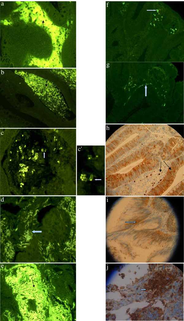
The study involved 14 patients (four with pre-malignant polyps and 10 with colorectal adenocarcinomas). The distribution of μ chains and CD15 markers varied in all of the biopsies, but delineated normal cell structures in the pre-malignant polyp specimens. B cell surface phenotype analysis of μ chain-positive cells identified a subset of CD79+/CD20-/CD21-/CD23- IPCs. The IPCs in certain areas showed the sporadic disintegration of inflammatory cell membranes or the accumulation of fluorescence in individual cells. IPC membrane disintegration was particularly marked in all of the adenocarcinoma samples, in which the CD15 markers also showed epithelial cell involvement. Furthermore, six of the ten adenocarcinoma samples had atypical and reorganised membranes that expressed an excess of both receptors and isolated small portions of tissue within the tumour.
The findings of this preliminary morphological study suggest the presence of membrane disintegration and remodelling mechanisms in the tumours. The newly-formed membranes expressed high concentrations of inflammatory cell receptors that can confer adhesive properties.
我们最近研究了炎症性肠病(IBD)患者炎症肠道组织样本中产生免疫球蛋白细胞(IPC)的定位,并确定了B淋巴细胞浸润的两种主要模式:一种以小B1细胞样IgM + / CD79 + / CD20 - / CD21 - / CD23 - / CD5 ± IPC的中度强基质定位为特征,另一种以具有不规则核的IPC的腺周定位为特征,这些IPC具有B细胞亚群特有的表面标志物(IgM和CD79),但其λ和κ链存在定量差异。对同一批患者还检测了CD15 + 受体,其定位于炎症细胞表面或肠上皮隐窝中。炎症组织中CD15 + 受体的分布仅限于细胞结构。本研究的目的是分析来自癌前息肉或腺癌患者的肠道活检标本中IPC和CD15 + 细胞形态或分布的变化。
使用多克隆山羊抗人μ链通过免疫荧光分析IPC。使用免疫过氧化物酶法检测癌前息肉标本的B细胞表面表型λ和κ链、CD79、CD20、CD21和CD23。使用免疫过氧化物酶法和单克隆抗CD15 IgM评估CD15 + 细胞。
该研究纳入了14例患者(4例癌前息肉患者和10例结直肠癌患者)。所有活检标本中μ链和CD15标志物的分布各不相同,但在癌前息肉标本中勾勒出正常细胞结构。对μ链阳性细胞的B细胞表面表型分析确定了一个CD79 + / CD20 - / CD21 - / CD23 - IPC亚群。某些区域的IPC显示出炎症细胞膜的散在崩解或单个细胞中荧光的积累。IPC膜崩解在所有腺癌样本中尤为明显,其中CD15标志物也显示上皮细胞受累。此外,10例腺癌样本中有6例具有非典型且重新组织的膜,其表达过量的两种受体并在肿瘤内分离出小部分组织。
这项初步形态学研究的结果表明肿瘤中存在膜崩解和重塑机制。新形成的膜表达高浓度的炎症细胞受体,可赋予黏附特性。