Cancer Therapeutics Program, Gene Regulation Laboratory, The Peter MacCallum Cancer Centre, St Andrews Place, East Melbourne 3002, Victoria, Australia.
Cell Death Dis. 2013 Feb 28;4(2):e519. doi: 10.1038/cddis.2013.9.
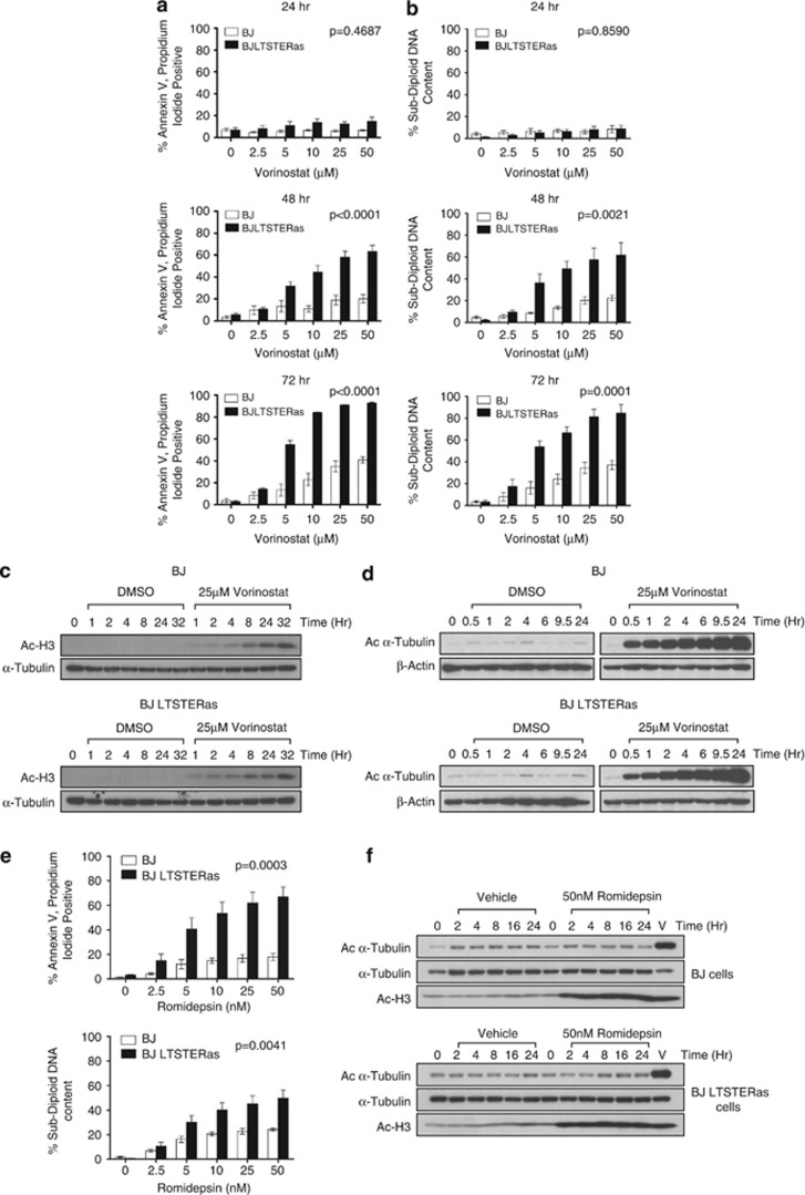
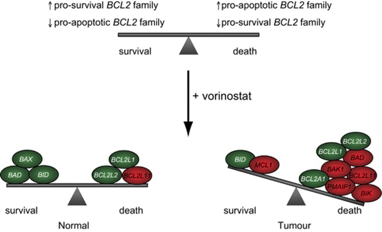
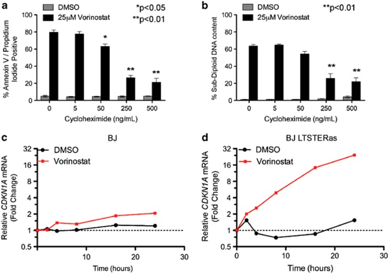
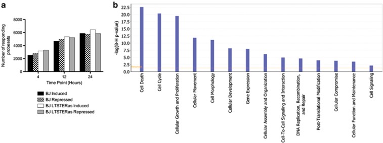
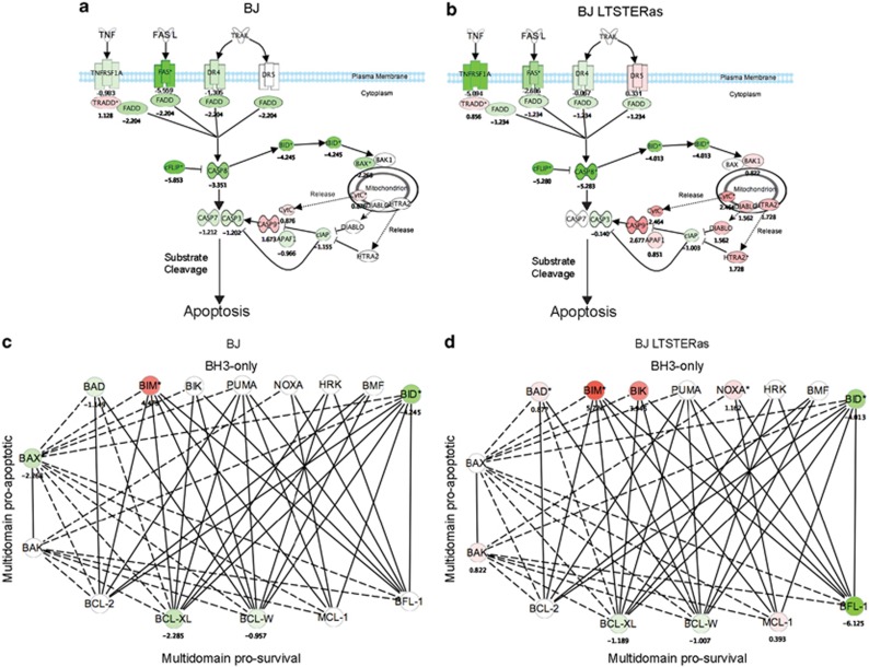
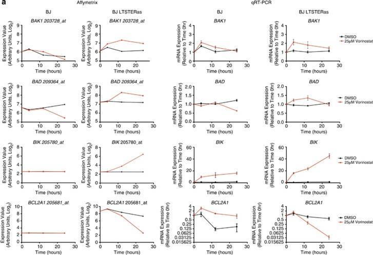
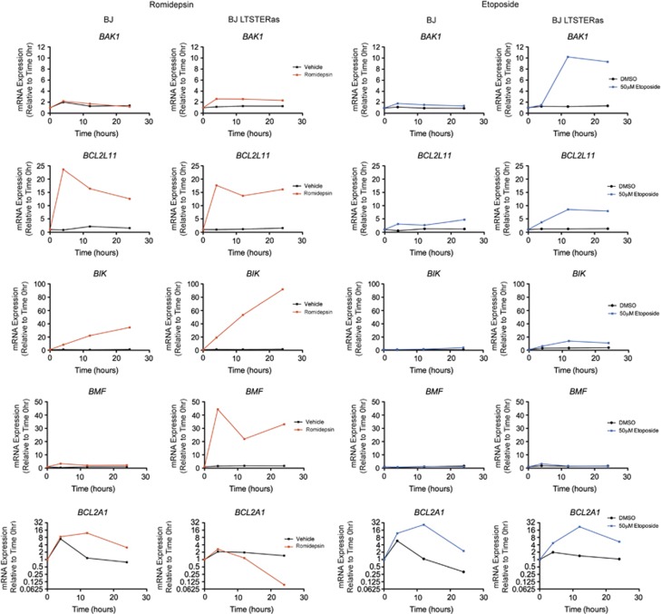
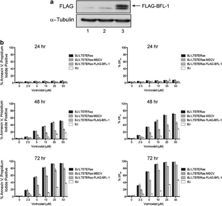
The identification of recurrent somatic mutations in genes encoding epigenetic enzymes has provided a strong rationale for the development of compounds that target the epigenome for the treatment of cancer. This notion is supported by biochemical studies demonstrating aberrant recruitment of epigenetic enzymes such as histone deacetylases (HDACs) and histone methyltransferases to promoter regions through association with oncogenic fusion proteins such as PML-RARα and AML1-ETO. HDAC inhibitors (HDACi) are potent inducers of tumor cell apoptosis; however, it remains unclear why tumor cells are more sensitive to HDACi-induced cell death than normal cells. Herein, we assessed the biological and molecular responses of isogenic normal and transformed cells to the FDA-approved HDACi vorinostat and romidepsin. Both HDACi selectively killed cells of diverse tissue origin that had been transformed through the serial introduction of different oncogenes. Time-course microarray expression profiling revealed that normal and transformed cells transcriptionally responded to vorinostat treatment. Over 4200 genes responded differently to vorinostat in normal and transformed cells and gene ontology and pathway analyses identified a tumor-cell-selective pro-apoptotic gene-expression signature that consisted of BCL2 family genes. In particular, HDACi induced tumor-cell-selective upregulation of the pro-apoptotic gene BMF and downregulation of the pro-survival gene BCL2A1 encoding BFL-1. Maintenance of BFL-1 levels in transformed cells through forced expression conferred vorinostat resistance, indicating that specific and selective engagement of the intrinsic apoptotic pathway underlies the tumor-cell-selective apoptotic activities of these agents. The ability of HDACi to affect the growth and survival of tumor cells whilst leaving normal cells relatively unharmed is fundamental to their successful clinical application. This study provides new insight into the transcriptional effects of HDACi in human donor-matched normal and transformed cells, and implicates specific molecules and pathways in the tumor-selective cytotoxic activity of these compounds.
鉴定编码表观遗传酶的基因中的复发性体细胞突变,为针对表观基因组开发治疗癌症的化合物提供了强有力的理论依据。这一概念得到了生化研究的支持,这些研究表明,表观遗传酶(如组蛋白去乙酰化酶 (HDAC) 和组蛋白甲基转移酶)通过与致癌融合蛋白(如 PML-RARα 和 AML1-ETO)结合,异常募集到启动子区域。HDAC 抑制剂 (HDACi) 是肿瘤细胞凋亡的有效诱导剂;然而,目前尚不清楚为什么肿瘤细胞比正常细胞对 HDACi 诱导的细胞死亡更敏感。在此,我们评估了同基因正常和转化细胞对 FDA 批准的 HDACi 伏立诺他和罗米地辛的生物学和分子反应。这两种 HDACi 选择性地杀死了通过连续引入不同致癌基因而转化的来自不同组织来源的细胞。时间过程微阵列表达谱分析显示,正常和转化细胞对伏立诺他治疗有转录反应。超过 4200 个基因对正常和转化细胞中的伏立诺他反应不同,基因本体论和途径分析确定了一个肿瘤细胞选择性促凋亡基因表达谱,该谱由 BCL2 家族基因组成。特别是,HDACi 诱导肿瘤细胞选择性地上调促凋亡基因 BMF 和下调促生存基因 BCL2A1 编码 BFL-1。通过强制表达在转化细胞中维持 BFL-1 水平赋予伏立诺他抗性,表明这些药物的肿瘤细胞选择性促凋亡活性的基础是内在凋亡途径的特异性和选择性参与。HDACi 能够影响肿瘤细胞的生长和存活,而使正常细胞相对不受伤害,这是其成功临床应用的基础。本研究提供了在人供体匹配的正常和转化细胞中 HDACi 的转录效应的新见解,并涉及这些化合物的肿瘤选择性细胞毒性活性的特定分子和途径。