Laboratory of Experimental Intensive Care and Anesthesiology, Academic Medical Center, Amsterdam, The Netherlands.
PLoS One. 2013;8(2):e57374. doi: 10.1371/journal.pone.0057374. Epub 2013 Feb 25.
Ventilator-induced lung injury (VILI) is characterized by vascular leakage and inflammatory responses eventually leading to pulmonary dysfunction. Vascular endothelial growth factor (VEGF) has been proposed to be involved in the pathogenesis of VILI. This study examines the inhibitory effect of dexamethasone on VEGF expression, inflammation and alveolar-capillary barrier dysfunction in an established murine model of VILI.
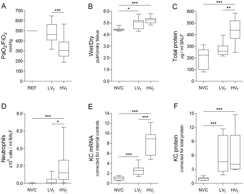
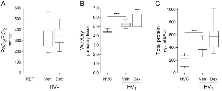
Healthy male C57Bl/6 mice were anesthetized, tracheotomized and mechanically ventilated for 5 hours with an inspiratory pressure of 10 cmH2O ("lower" tidal volumes of ∼7.5 ml/kg; LVT) or 18 cmH2O ("higher" tidal volumes of ∼15 ml/kg; HVT). Dexamethasone was intravenously administered at the initiation of HVT-ventilation. Non-ventilated mice served as controls. Study endpoints included VEGF and inflammatory mediator expression in lung tissue, neutrophil and protein levels in bronchoalveolar lavage fluid, PaO2 to FiO2 ratios and lung wet to dry ratios.
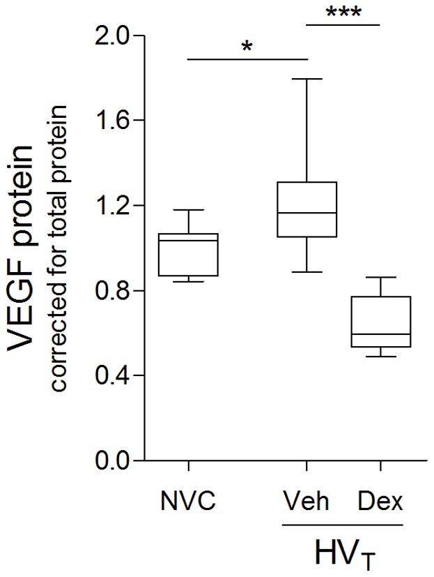
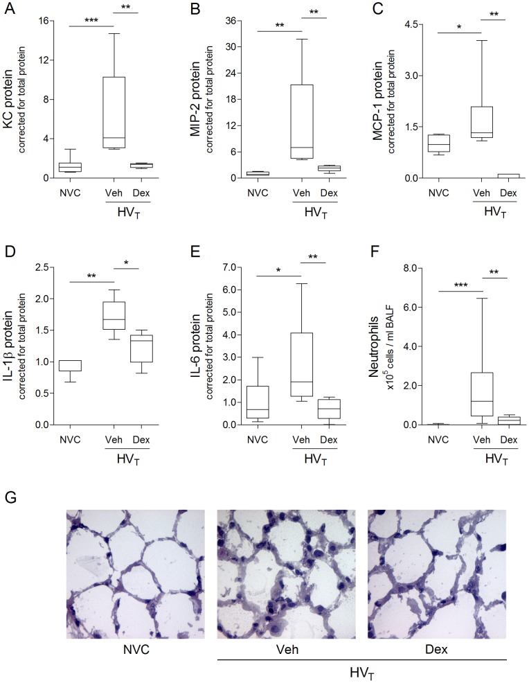
Particularly HVT-ventilation led to alveolar-capillary barrier dysfunction as reflected by reduced PaO2 to FiO2 ratios, elevated alveolar protein levels and increased lung wet to dry ratios. Moreover, VILI was associated with enhanced VEGF production, inflammatory mediator expression and neutrophil infiltration. Dexamethasone treatment inhibited VEGF and pro-inflammatory response in lungs of HVT-ventilated mice, without improving alveolar-capillary permeability, gas exchange and pulmonary edema formation.
Dexamethasone treatment completely abolishes ventilator-induced VEGF expression and inflammation. However, dexamethasone does not protect against alveolar-capillary barrier dysfunction in an established murine model of VILI.
呼吸机相关性肺损伤(VILI)的特征是血管渗漏和炎症反应,最终导致肺功能障碍。血管内皮生长因子(VEGF)被认为参与了 VILI 的发病机制。本研究在建立的 VILI 小鼠模型中,研究了地塞米松对 VEGF 表达、炎症和肺泡毛细血管屏障功能障碍的抑制作用。
健康雄性 C57Bl/6 小鼠接受麻醉、气管切开和机械通气 5 小时,吸气压力为 10 cmH2O(“低”潮气量约 7.5 ml/kg;LVT)或 18 cmH2O(“高”潮气量约 15 ml/kg;HVT)。在开始 HVT 通气时静脉给予地塞米松。未通气的小鼠作为对照。研究终点包括肺组织中 VEGF 和炎症介质的表达、支气管肺泡灌洗液中的中性粒细胞和蛋白水平、PaO2 与 FiO2 的比值以及肺湿干比。
特别是 HVT 通气导致肺泡毛细血管屏障功能障碍,表现为 PaO2 与 FiO2 的比值降低、肺泡蛋白水平升高和肺湿干比增加。此外,VILI 与 VEGF 产生增加、炎症介质表达和中性粒细胞浸润有关。地塞米松治疗抑制了 HVT 通气小鼠肺部的 VEGF 和促炎反应,但不能改善肺泡毛细血管通透性、气体交换和肺水肿形成。
地塞米松治疗完全抑制了呼吸机相关性 VEGF 表达和炎症反应。然而,地塞米松不能预防已建立的 VILI 小鼠模型中的肺泡毛细血管屏障功能障碍。