Department of Pharmacology and Experimental Therapeutics, Louisiana State University Health Sciences Center, New Orleans, Louisiana, United States of America.
PLoS One. 2013;8(2):e57805. doi: 10.1371/journal.pone.0057805. Epub 2013 Feb 25.
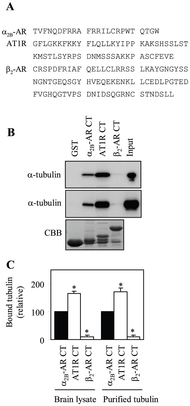
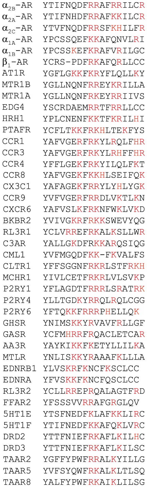
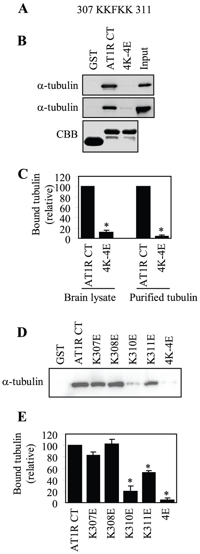
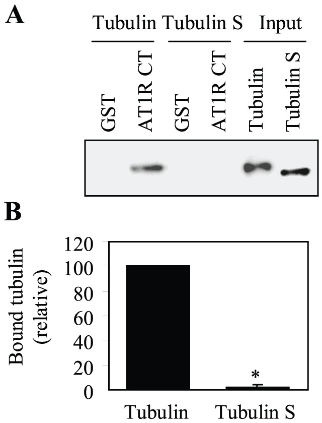
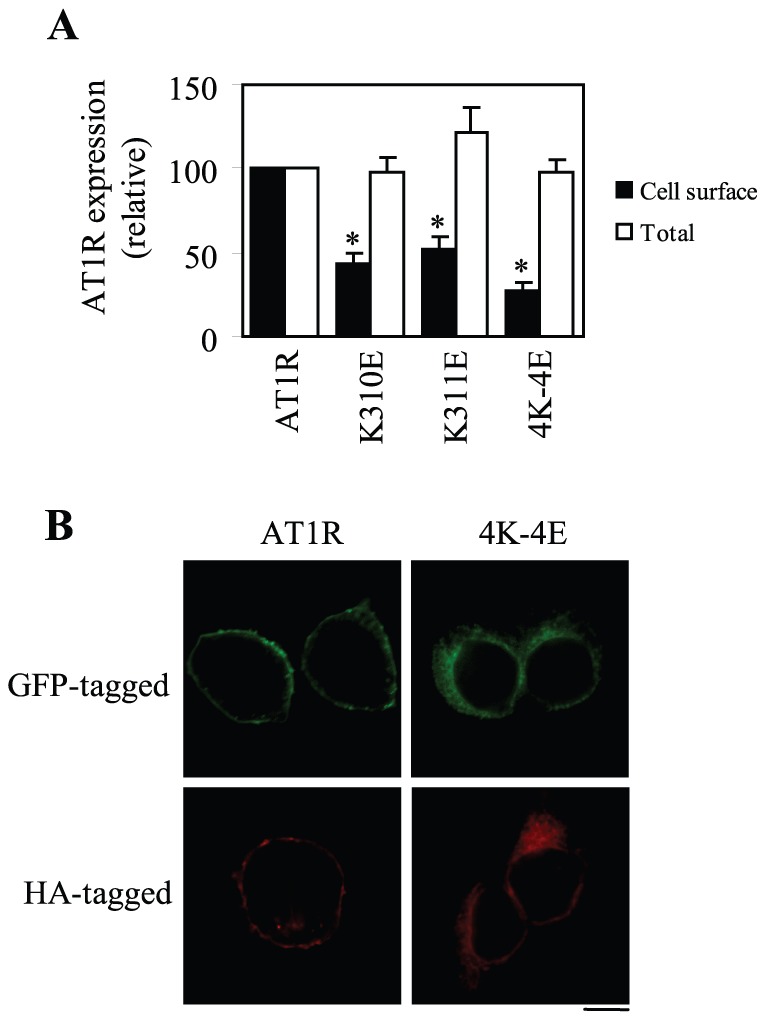
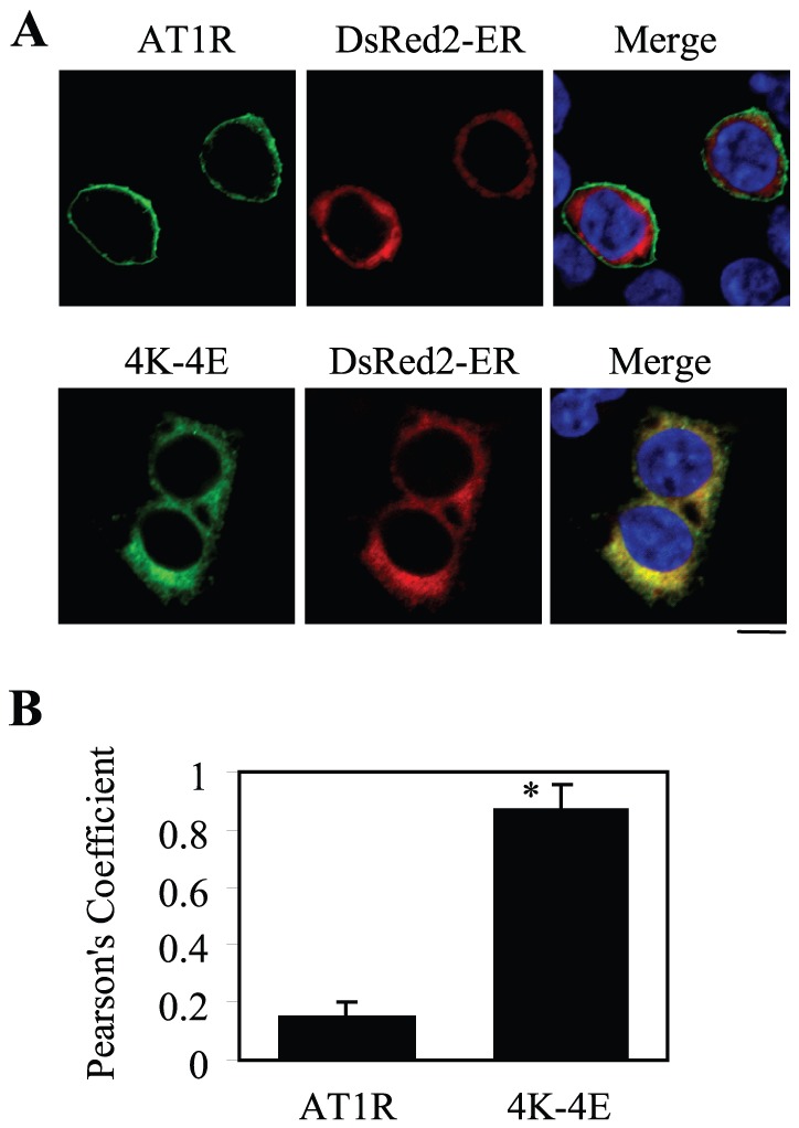
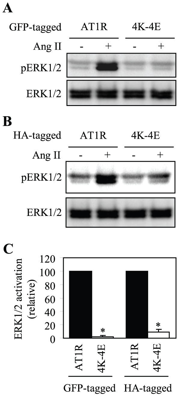
The physiological and pathological functions of angiotensin II are largely mediated through activating the cell surface angiotensin II type 1 receptor (AT1R). However, the molecular mechanisms underlying the transport of newly synthesized AT1R from the endoplasmic reticulum (ER) to the cell surface remain poorly defined. Here we demonstrated that the C-terminus (CT) of AT1R directly and strongly bound to tubulin and the binding domains were mapped to two consecutive Lys residues at positions 310 and 311 in the CT membrane-proximal region of AT1R and the acidic CT of tubulin, suggestive of essentially ionic interactions between AT1R and tubulin. Furthermore, mutation to disrupt tubulin binding dramatically inhibited the cell surface expression of AT1R, arrested AT1R in the ER, and attenuated AT1R-mediated signaling measured as ERK1/2 activation. These data demonstrate for the first time that specific Lys residues in the CT juxtamembrane region regulate the processing of AT1R through interacting with tubulin. These data also suggest an important role of the microtubule network in the cell surface transport of AT1R.
血管紧张素 II 的生理和病理功能主要通过激活细胞表面的血管紧张素 II 型 1 受体(AT1R)来介导。然而,新合成的 AT1R 从内质网(ER)向细胞表面转运的分子机制仍未完全确定。在这里,我们证明 AT1R 的 C 端(CT)直接并强烈结合微管蛋白,并且结合结构域被映射到 AT1R 的 CT 膜近端区域中的两个连续赖氨酸残基 310 和 311 以及微管蛋白的酸性 CT,提示 AT1R 和微管蛋白之间存在本质上的离子相互作用。此外,突变以破坏微管蛋白结合可显著抑制 AT1R 的细胞表面表达,将 AT1R 阻滞在 ER 中,并减弱 AT1R 介导的信号转导,如 ERK1/2 激活。这些数据首次表明,CT 近膜区的特定赖氨酸残基通过与微管蛋白相互作用调节 AT1R 的加工。这些数据还表明微管网络在 AT1R 的细胞表面转运中起着重要作用。