Department of Anatomy I, University of Cologne, Faculty of Medicine and University Hospital Cologne, Kerpener Str. 62, 50937, Cologne, Germany.
Institute II of Pharmacology, Center of Pharmacology, University of Cologne, Faculty of Medicine and University Hospital Cologne, Gleueler Str. 24, 50931, Cologne, Germany.
Histochem Cell Biol. 2022 May;157(5):513-524. doi: 10.1007/s00418-022-02087-z. Epub 2022 Feb 28.
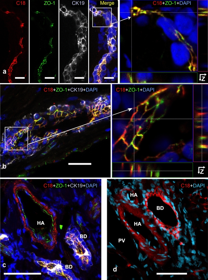
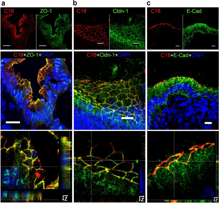
Animal models and clinical studies suggest an influence of angiotensin II (AngII) on the pathogenesis of liver diseases via the renin-angiotensin system. AngII application increases portal blood pressure, reduces bile flow, and increases permeability of liver tight junctions. Establishing the subcellular localization of angiotensin II receptor type 1 (AT1R), the main AngII receptor, helps to understand the effects of AngII on the liver. We localized AT1R in situ in human and porcine liver and porcine gallbladder by immunohistochemistry. In order to do so, we characterized commercial anti-AT1R antibodies regarding their capability to recognize heterologous human AT1R in immunocytochemistry and on western blots, and to detect AT1R using overlap studies and AT1R-specific blocking peptides. In hepatocytes and canals of Hering, AT1R displayed a tram-track-like distribution, while in cholangiocytes AT1R appeared in a honeycomb-like pattern; i.e., in liver epithelia, AT1R showed an equivalent distribution to that in the apical junctional network, which seals bile canaliculi and bile ducts along the blood-bile barrier. In intrahepatic blood vessels, AT1R was most prominent in the tunica media. We confirmed AT1R localization in situ to the plasma membrane domain, particularly between tight and adherens junctions in both human and porcine hepatocytes, cholangiocytes, and gallbladder epithelial cells using different anti-AT1R antibodies. Localization of AT1R at the junctional complex could explain previously reported AngII effects and predestines AT1R as a transmitter of tight junction permeability.
动物模型和临床研究表明,血管紧张素 II(AngII)通过肾素-血管紧张素系统对肝脏疾病的发病机制有影响。AngII 的应用会增加门静脉血压,减少胆汁流量,并增加肝紧密连接的通透性。确定血管紧张素 II 受体 1(AT1R)的亚细胞定位,即主要的 AngII 受体,有助于了解 AngII 对肝脏的影响。我们通过免疫组织化学方法原位定位了人肝、猪肝和猪胆囊中的 AT1R。为此,我们对商业抗 AT1R 抗体进行了特征描述,以确定其在免疫细胞化学和 Western blot 中识别异源人 AT1R 的能力,以及使用重叠研究和 AT1R 特异性阻断肽检测 AT1R 的能力。在肝细胞和 Hering 胆管中,AT1R 呈 tram-track 样分布,而在胆管细胞中,AT1R 呈蜂窝状分布;即在肝上皮细胞中,AT1R 的分布与顶端连接网络相当,该网络沿着血-胆屏障密封胆小管和胆管。在肝内血管中,AT1R 在中膜最为突出。我们使用不同的抗 AT1R 抗体证实了 AT1R 原位定位于质膜域,特别是在人肝和猪肝、胆管和胆囊上皮细胞的紧密连接和黏附连接之间。AT1R 在连接复合体中的定位可以解释之前报道的 AngII 作用,并使 AT1R 成为紧密连接通透性的递质。