Department of Chemical and Biomolecular Engineering, University of Illinois, 600 S. Mathews Avenue, Urbana, IL 61801, USA.
Biomaterials. 2013 May;34(15):3902-11. doi: 10.1016/j.biomaterials.2013.02.015. Epub 2013 Feb 27.
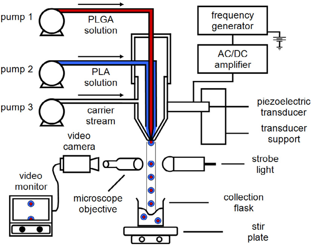
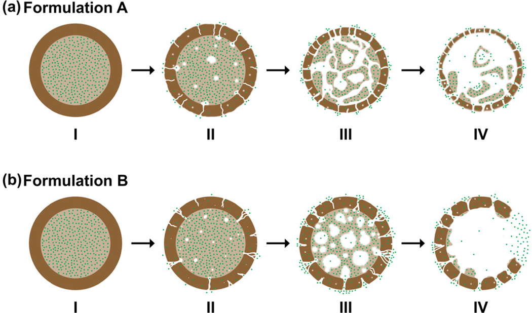
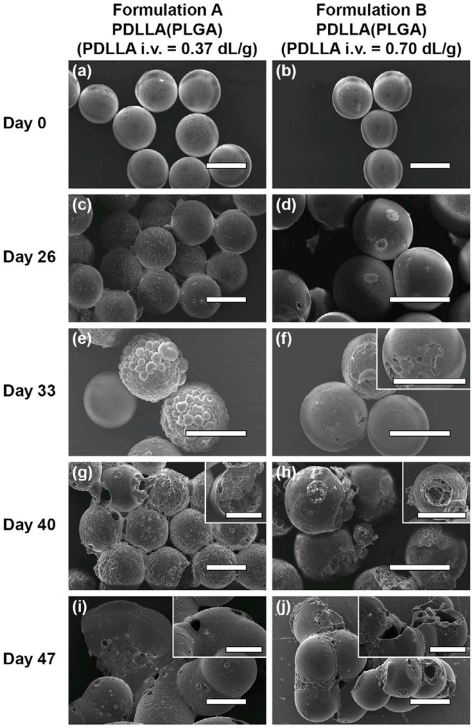
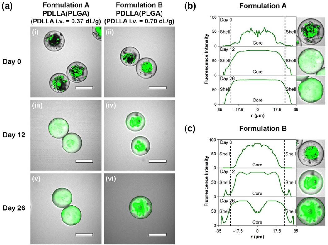
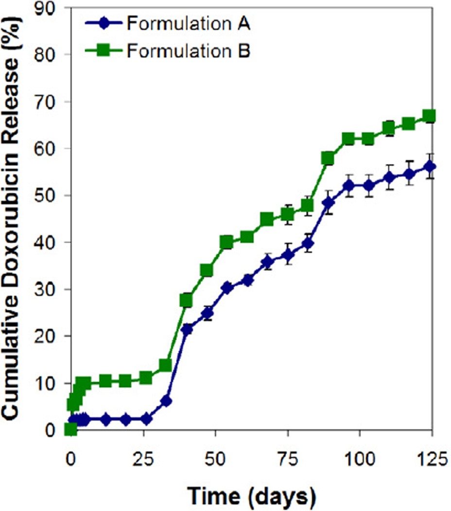
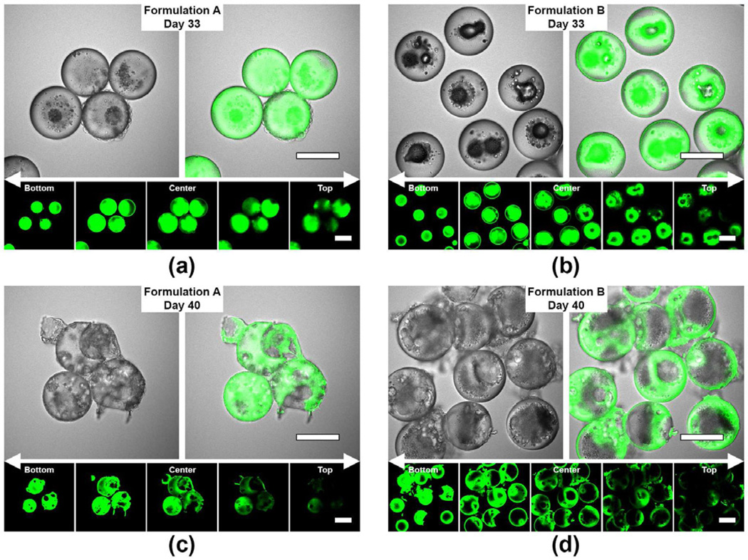
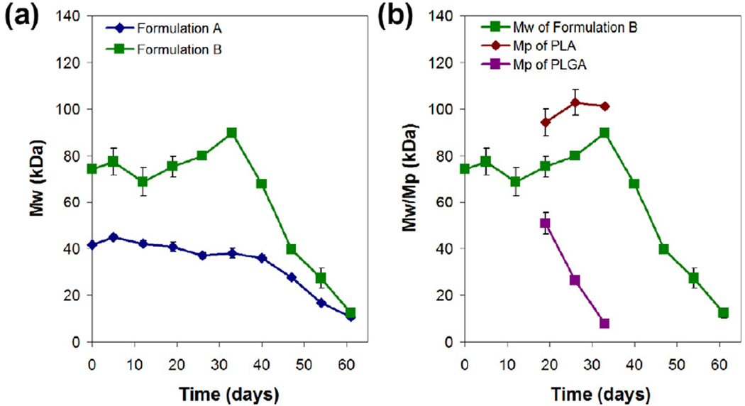
The drug release and degradation behavior of two double-walled microsphere formulations consisting of a doxorubicin-loaded poly(d,l-lactic-co-glycolic acid) (PLGA) core (∼46 kDa) surrounded by a poly(d,l-lactic acid) (PDLLA) shell layer (∼55 and 116 kDa) were examined. It was postulated that different molecular weights of the shell layer could modulate the erosion of the outer coating and limit the occurrence of water penetration into the inner drug-loaded core on various time scales, and therefore control the drug release from the microspheres. For both microsphere formulations, the drug release profiles were observed to be similar. The degradation of the microspheres was monitored for a period of about nine weeks and analyzed using scanning electron microscopy, laser scanning confocal microscopy, and gel permeation chromatography. Interestingly, both microsphere formulations exhibited occurrence of bulk erosion of PDLLA on a similar time scale despite different PDLLA molecular weights forming the shell layer. The shell layer of the double-walled microspheres served as an effective diffusion barrier during the initial lag phase period and controlled the release rate of the hydrophilic drug independent of the molecular weight of the shell layer.
两种双层微球制剂的药物释放和降解行为进行了研究,这两种双层微球制剂由包载阿霉素的聚(丙交酯-乙交酯)(PLGA)核(46 kDa)和聚(丙交酯)(PDLLA)壳层(55 和 116 kDa)组成。据推测,不同分子量的壳层可以调节外层涂层的侵蚀,并限制水在不同时间尺度上渗透到载药的内核中,从而控制微球的药物释放。对于这两种微球制剂,药物释放曲线相似。微球的降解在大约九周的时间内进行监测,并使用扫描电子显微镜、激光共聚焦显微镜和凝胶渗透色谱进行分析。有趣的是,尽管形成壳层的 PDLLA 分子量不同,但两种微球制剂均表现出 PDLLA 的整体侵蚀发生在相似的时间范围内。双层微球的壳层在初始滞后阶段充当有效的扩散屏障,并控制亲水性药物的释放速率,而与壳层的分子量无关。