Division of Orthopedics, Shriners Hospital for Children, Montréal, Quebec, Canada.
PLoS One. 2013;8(2):e56790. doi: 10.1371/journal.pone.0056790. Epub 2013 Feb 15.
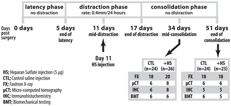
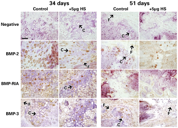
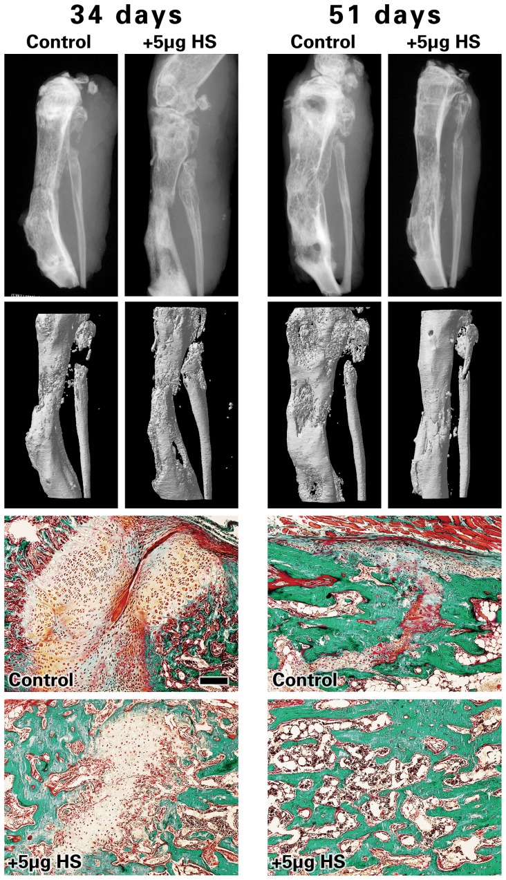
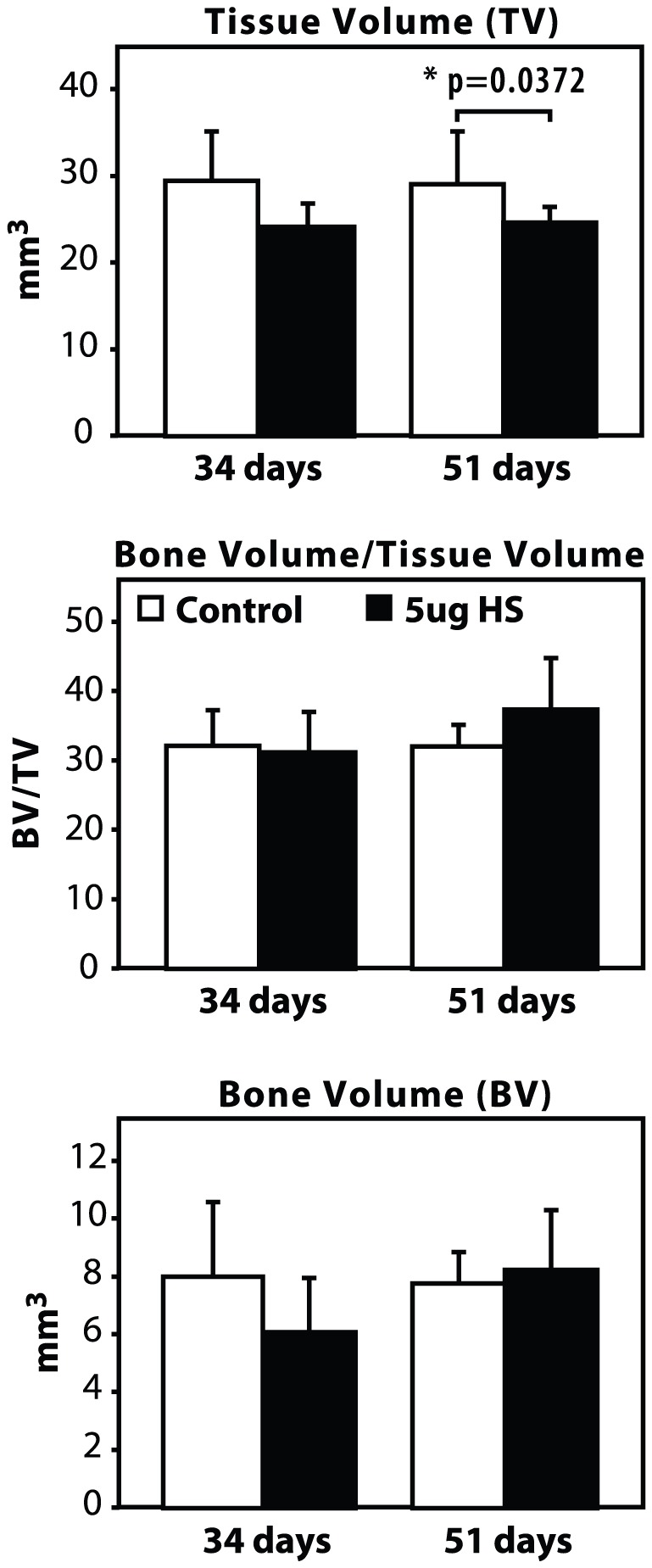
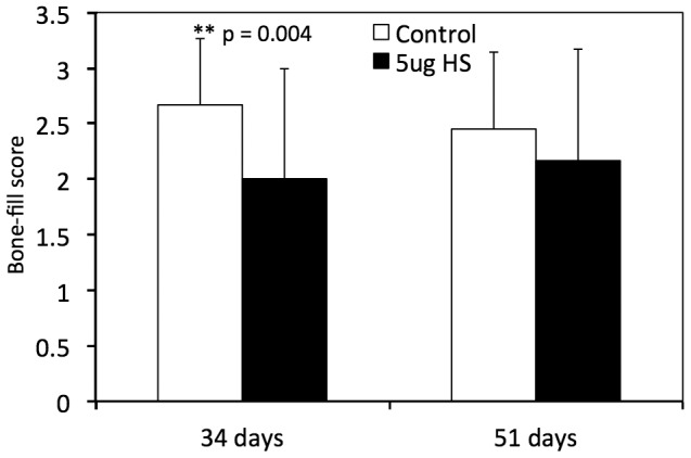
Bone morphogenetic proteins (BMPs) are recognized for their ability to induce bone formation in vivo and in vitro. Their osteogenic and osteoinductive properties are tightly regulated by the secretion of specific BMP antagonists, which have been shown to physically bind and sometimes be blocked by the extracellular proteoglycan heparan sulphate side chains (from hereon referred to as HS). The purpose of this study was to investigate if local application of 5 µg of HS proteoglycan to a bone regenerate site in a mouse model of distraction osteogenesis (DO) can accelerate bone healing and affect the expression of key members of the BMP signaling pathway. DO was performed on the right tibia of 115 adult male wild-type mice. At mid-distraction (day 11), half the group was injected locally with 5 µg of HS, while the other half was injected with saline. The mice were sacrificed at 2 time-points: mid-consolidation (34 days) and full consolidation (51 days). The distracted tibial zone was then collected for analysis by μCT, radiology, biomechanical testing, immunohistochemistry, and histology. While μCT data showed no statistically significant difference in bone formation, the results of biomechanical testing in stiffness and ultimate force were significantly lower in the HS-injected bones at 51 days, compared to controls. Immunohistochemistry results also suggested a decrease in expression of several key members of the BMP signaling pathway at 34 days. Furthermore, wound dehiscence and infection rates were significantly elevated in the HS group compared to the controls, which resulted in a higher rate of euthanasia in the treatment group. Our findings demonstrate that exogenous application of 5 µg of HS in the distracted gap of a murine model had a negative impact on bone and wound healing.
骨形态发生蛋白(BMPs)因其在体内和体外诱导骨形成的能力而被认可。它们的成骨和诱导成骨特性受到特定 BMP 拮抗剂的分泌的严格调节,这些拮抗剂已被证明可以物理结合并有时被细胞外蛋白聚糖硫酸乙酰肝素侧链(此后简称 HS)阻断。本研究旨在探讨在牵张成骨(DO)的小鼠模型中,将 5µg 的 HS 蛋白聚糖局部应用于骨再生部位是否可以加速骨愈合并影响 BMP 信号通路关键成员的表达。DO 在 115 只成年雄性野生型小鼠的右侧胫骨上进行。在中期牵张(第 11 天)时,一半的小鼠局部注射 5µg 的 HS,另一半注射生理盐水。在两个时间点处死小鼠:中期骨整合(34 天)和完全骨整合(51 天)。然后收集牵张的胫骨区域进行 μCT、放射学、生物力学测试、免疫组织化学和组织学分析。虽然 μCT 数据显示骨形成没有统计学上的显著差异,但在 51 天时,生物力学测试中刚度和最大力的结果显示,注射 HS 的骨骼明显低于对照组。免疫组织化学结果还表明,在 34 天时,BMP 信号通路的几个关键成员的表达减少。此外,与对照组相比,HS 组的伤口裂开和感染率显著升高,这导致治疗组的安乐死率更高。我们的研究结果表明,在小鼠模型的牵张间隙中应用 5µg 的外源性 HS 对骨和伤口愈合有负面影响。