Carman and Ann Adams Department of Pediatrics, Wayne State University School of Medicine, Detroit, Michigan, USA; Department of Neurology, Wayne State University School of Medicine, Detroit, Michigan, USA; Translational Imaging Laboratory, Children's Hospital of Michigan, Detroit, Michigan, USA.
J Magn Reson Imaging. 2013 Nov;38(5):1152-61. doi: 10.1002/jmri.24076. Epub 2013 Mar 5.
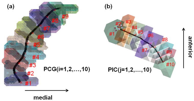
To explore whether diffusion-weighted imaging (DWI) can localize specific segments of primary motor areas in children with Sturge-Weber syndrome (SWS), this study investigated the corticospinal tract (CST) between precentral gyrus (PCG) and posterior limb of internal capsule (PIC).
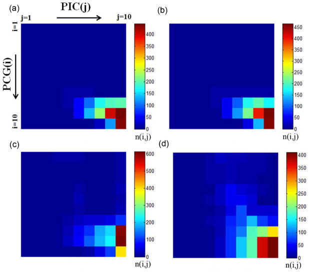
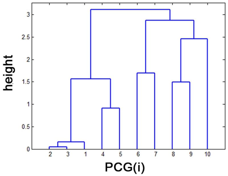
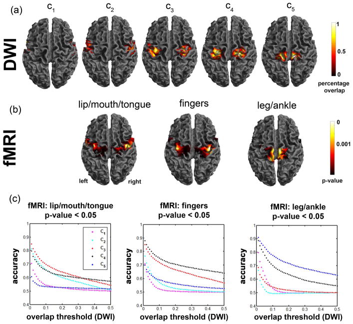
DWI was performed on 32 healthy children and seven children with unilateral SWS affecting the sensorimotor area variably. A hierarchical dendrogram was applied to find PCG-segments uniquely connected to PIC-segments. The resulting PCG-clusters were used to image primary motor pathways in DWI and find metabolic abnormalities of primary motor areas in positron emission tomography (PET) scans.
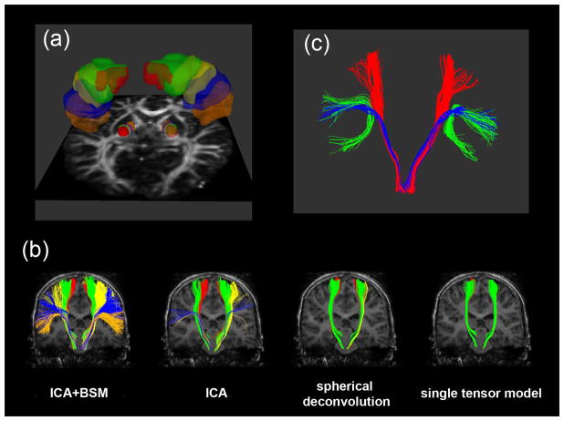
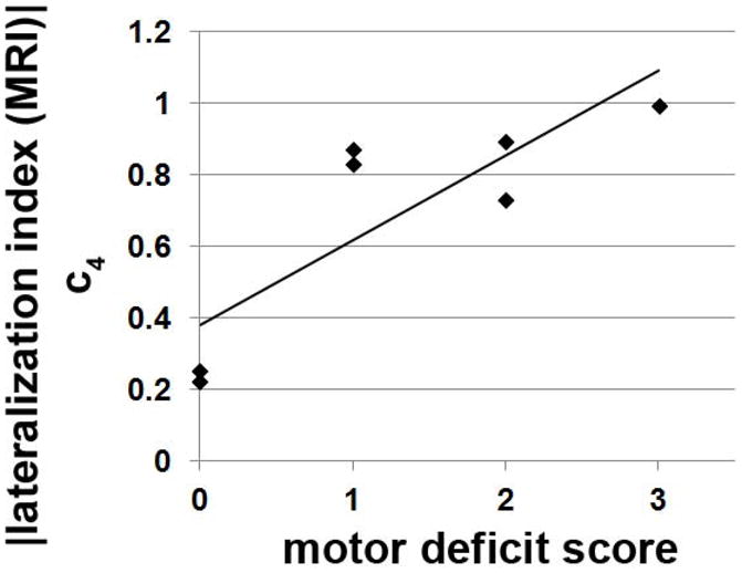
In healthy children, five PCG-clusters were found to have unique CST courses, corresponding to CST segments of mouth/lip, fingers, and leg/ankle primary motor areas determined by functional magnetic resonance imaging (fMRI). In children with SWS, reduced streamlines in these PCG clusters were highly correlated with glucose-hypometabolism on PET (R(2) = 0.2312, P = 0.0032). Impaired CST segment corresponding to finger movements correlated with severity of hand motor deficit.
The presented method can detect impaired CST segments corresponding to specific motor functions in young children who cannot cooperate for fMRI. This approach can be clinically useful for a noninvasive presurgical evaluation of cortical motor areas in such children.
本研究旨在探讨弥散加权成像(DWI)是否可以定位 Sturge-Weber 综合征(SWS)患儿初级运动区的特定节段,以研究中央前回(PCG)与内囊后肢(PIC)之间的皮质脊髓束(CST)。
对 32 名健康儿童和 7 名单侧 SWS 患儿进行了 DWI 检查,这些患儿的感觉运动区受到不同程度的影响。采用层次聚类树图来寻找唯一连接 PIC 节段的 PCG 节段。由此产生的 PCG 聚类被用于 DWI 中初级运动通路的成像,并在正电子发射断层扫描(PET)中寻找初级运动区的代谢异常。
在健康儿童中,发现了五个具有独特 CST 轨迹的 PCG 聚类,与功能磁共振成像(fMRI)确定的口/唇、手指和腿/脚踝初级运动区的 CST 节段相对应。在 SWS 患儿中,这些 PCG 聚类中减少的流线与 PET 上的葡萄糖代谢低下高度相关(R²=0.2312,P=0.0032)。对应手指运动的 CST 节段受损与手部运动缺陷的严重程度相关。
该方法可检测出无法进行 fMRI 检查的幼儿中对应特定运动功能的受损 CST 节段。这种方法可在这些儿童的皮质运动区进行非侵入性术前评估方面具有临床应用价值。