Carman and Ann Adams Department of Pediatrics, School of Medicine, Wayne State University, Detroit, Michigan 48201, USA.
Epilepsia. 2013 Aug;54(8):1381-90. doi: 10.1111/epi.12199. Epub 2013 Jun 17.
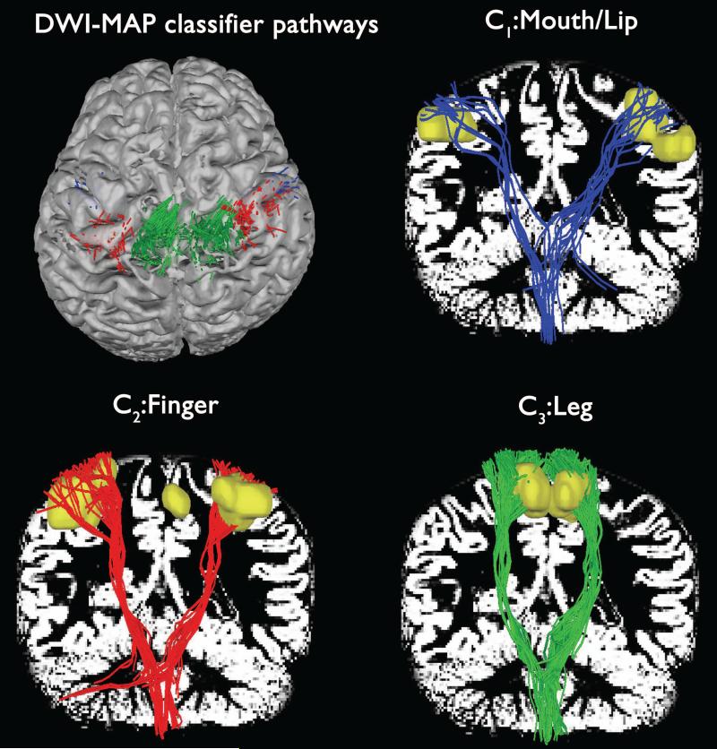
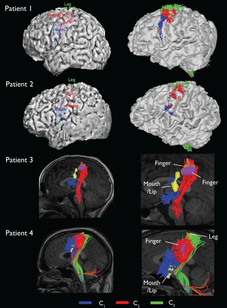
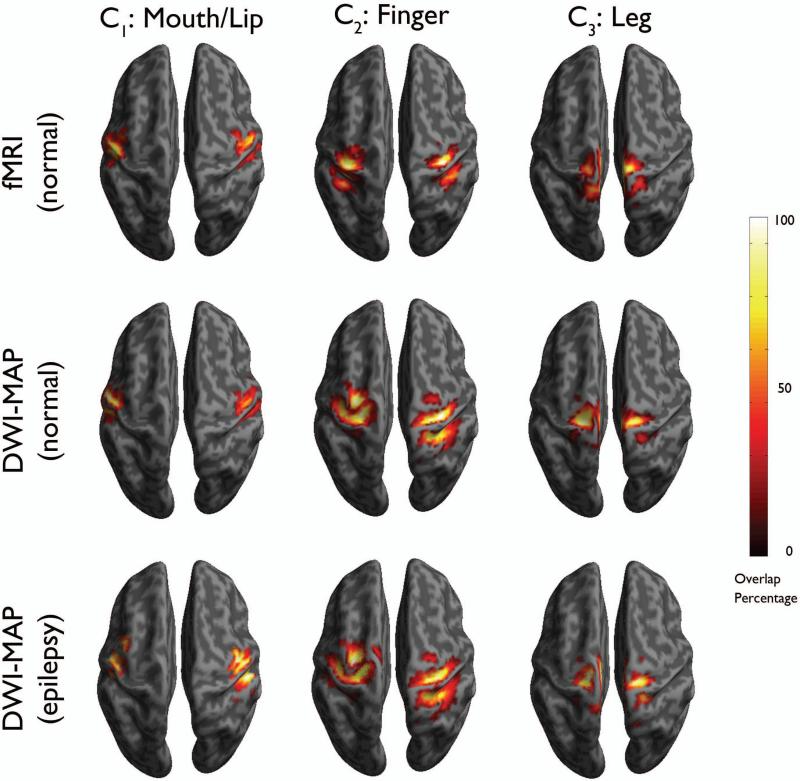
As an alternative tool to identify cortical motor areas for planning surgical resection in children with focal epilepsy, the present study proposed a maximum a posteriori probability (MAP) classification of corticospinal tract (CST) visualized by diffusion MR tractography.
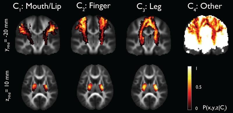
Diffusion-weighted imaging (DWI) was performed in 17 normally developing children and 20 children with focal epilepsy. An independent component analysis tractography combined with ball-stick model was performed to identify unique CST pathways originating from mouth/lip, finger, and leg areas determined by functional magnetic resonance imaging (fMRI) in healthy children and electrical stimulation mapping (ESM) in children with epilepsy. Group analyses were performed to construct stereotaxic probability maps of primary motor pathways connecting precentral gyrus and posterior limb of internal capsule, and then utilized to design a novel MAP classifier that can sort individual CST fibers associated with three classes of interest: mouth/lip, fingers, and leg. A systematic leave-one-out approach was applied to train an optimal classifier. A match was considered to occur if classified fibers contacted or surrounded true areas localized by fMRI and ESM.
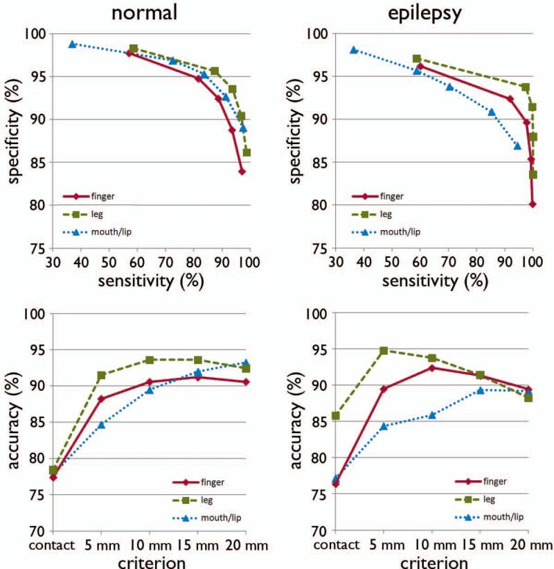
It was found that the DWI-MAP provided high accuracy for the CST fibers terminating in proximity to the localization of fMRI/ESM: 78%/77% for mouth/lip, 77%/76% for fingers, 78%/86% for leg (contact), and 93%/89% for mouth/lip, 91%/89% for fingers, and 92%/88% for leg (surrounded within 2 cm).
This study provides preliminary evidence that in the absence of fMRI and ESM data, the DWI-MAP approach can effectively retrieve the locations of cortical motor areas and underlying CST courses for planning epilepsy surgery.
作为识别儿童局灶性癫痫手术切除皮质运动区的替代工具,本研究提出了一种基于扩散磁共振束追踪技术的皮质脊髓束(CST)最大后验概率(MAP)分类方法。
对 17 名正常发育儿童和 20 名局灶性癫痫儿童进行弥散加权成像(DWI)。采用独立成分分析束追踪技术与球棒模型相结合,以识别源自健康儿童功能磁共振成像(fMRI)和癫痫儿童电刺激映射(ESM)确定的口/唇、手指和腿部区域的独特 CST 通路。进行组分析以构建连接中央前回和内囊后肢的主要运动通路的立体定向概率图,然后利用该概率图设计一种新的 MAP 分类器,可对与三个感兴趣类别(口/唇、手指和腿部)相关的个体 CST 纤维进行分类。采用系统的留一法进行最优分类器的训练。如果分类纤维与 fMRI 和 ESM 定位的真实区域接触或包围,则认为匹配发生。
发现 DWI-MAP 对 CST 纤维的定位具有很高的准确性,这些纤维与 fMRI/ESM 定位接近:口/唇为 78%/77%,手指为 77%/76%,腿部(接触)为 78%/86%,口/唇为 93%/89%,手指为 91%/89%,腿部(包围在 2cm 内)为 92%/88%。
本研究初步证明,在缺乏 fMRI 和 ESM 数据的情况下,DWI-MAP 方法可以有效地检索皮质运动区的位置及其下 CST 轨迹,以规划癫痫手术。