Division of Gastroenterology and Liver Disease, University Hospitals of Cleveland, Cleveland, Ohio, USA.
Clin Transl Gastroenterol. 2013 Mar 7;4(3):e31. doi: 10.1038/ctg.2013.2.
Obesity-associated carcinogenesis is postulated to be mediated through the proliferative actions of insulin and the insulin-like growth factor (IGF) family. The aim of this study was to determine whether the insulin/IGF-1 pathway is involved in the sequential progression from metaplastic Barrett's esophagus (BE) to dysplasia to esophageal adenocarcinoma (EAC).
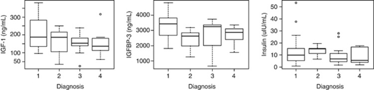
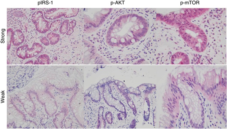
Fasting serum levels of insulin, glucose, IGF-1, insulin growth factor binding protein-1 (IGFBP1), and IGFBP3 were measured in 44 non-dysplastic, 9 low-grade dysplasia (LGD), 12 high-grade dysplasia (HGD), and 10 EAC subjects. Immunohistochemistry was performed on paraffin-embedded tissue derived from BE cases using rabbit monoclonal antibodies to p-mammalian target of rapamycin (mTOR) and p-AKT, mouse monoclonal antibody to Ki-67, and rabbit polyclonal antibody to p-insulin receptor substrate 1 (IRS1).
Nineteen of 44 (43.2%) BE, 5/9 (55%) LGD, 8/12 (66.7%) HGD and EAC 7/10 (70%) cases showed strong staining for p-IRS1. A significantly higher proportion of HGD/EAC subjects showed p-IRS1 staining when compared with BE/LGD subjects, 63.6% vs. 41.5%, P<0.05. p-IRS1 immunostaining was moderately correlated with strong immunostaining of the downstream mediators p-AKT and p-mTOR (Spearman correlation coefficient=0.167 and 0.27 for p-IRS1/p-AKT and for p-IRS1/p-mTOR, respectively) and the proliferation marker Ki-67 (Spearman correlation coefficient=0.20, P=0.09). However, systemic levels of insulin, IGF-1, or IGF-2 were not associated with tissue immunostaining of p-IRS1.
Activation of the insulin/IGF-1 pathway in BE may be associated with cellular proliferation and appears to have a role in the progression from metaplasia to cancer. The activation of the insulin/IGF-1 pathway at the tissue level is likely complex and does not have a simple association with systemic measures of insulin or IGF-1.
肥胖相关的致癌作用被认为是通过胰岛素和胰岛素样生长因子(IGF)家族的增殖作用介导的。本研究的目的是确定胰岛素/IGF-1 通路是否参与从化生的 Barrett 食管(BE)到异型增生到食管腺癌(EAC)的连续进展。
在 44 例非异型增生、9 例低级别异型增生(LGD)、12 例高级别异型增生(HGD)和 10 例 EAC 患者中测量空腹血清胰岛素、葡萄糖、IGF-1、胰岛素生长因子结合蛋白-1(IGFBP1)和 IGFBP3 的水平。使用兔单克隆抗体针对哺乳动物雷帕霉素靶蛋白(mTOR)和 p-AKT、鼠单克隆抗体针对 Ki-67 以及兔多克隆抗体针对 p-胰岛素受体底物 1(IRS1)对 BE 病例的石蜡包埋组织进行免疫组织化学染色。
在 44 例 BE 中有 19 例(43.2%)、9 例 LGD 中有 5 例(55%)、12 例 HGD 中有 8 例(66.7%)和 10 例 EAC 中有 7 例(70%)表现出强烈的 p-IRS1 染色。HGD/EAC 患者中 p-IRS1 染色的比例明显高于 BE/LGD 患者,分别为 63.6%和 41.5%,P<0.05。p-IRS1 免疫染色与下游介质 p-AKT 和 p-mTOR 的强免疫染色(Spearman 相关系数分别为 0.167 和 0.27)和增殖标志物 Ki-67(Spearman 相关系数为 0.20,P=0.09)中度相关。然而,胰岛素、IGF-1 或 IGF-2 的系统水平与 p-IRS1 的组织免疫染色无关。
BE 中胰岛素/IGF-1 通路的激活可能与细胞增殖有关,并可能在化生到癌症的进展中发挥作用。组织水平的胰岛素/IGF-1 通路的激活可能很复杂,并且与胰岛素或 IGF-1 的系统测量没有简单的关联。