Soft Tissue Biophysics Laboratory, Department of Chemical Engineering, McGill University, Montreal, QC H3A 2B2, Canada.
J Cell Mol Med. 2013 Apr;17(4):508-17. doi: 10.1111/jcmm.12034. Epub 2013 Mar 11.
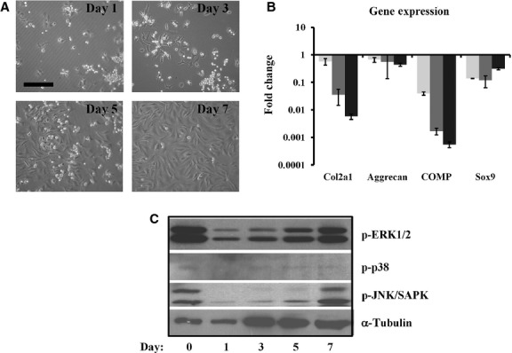
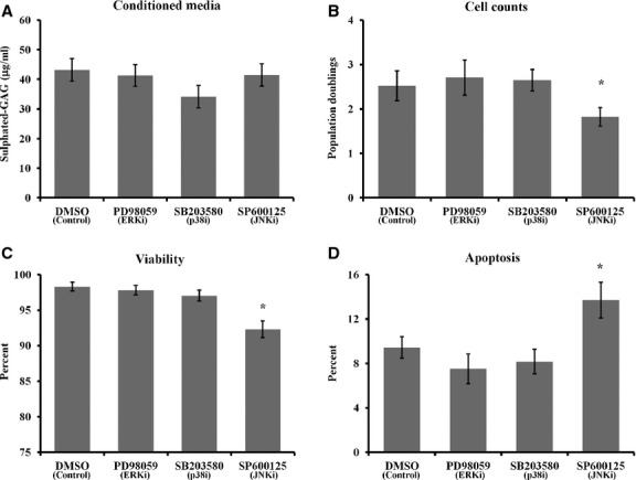
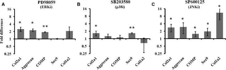
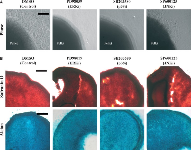
Articular cartilage is an avascular tissue with poor regenerative capacity following injury, a contributing factor to joint degenerative disease. Cell-based therapies for cartilage tissue regeneration have rapidly advanced; however, expansion of autologous chondrocytes in vitro using standard methods causes 'dedifferentiation' into fibroblastic cells. Mitogen-activated protein kinase (MAPK) signalling is crucial for chondrocyte metabolism and matrix production, and changes in MAPK signals can affect the phenotype of cultured cells. We investigated the effects of inhibition of MAPK signalling on chondrocyte dedifferentiation during monolayer culture. Blockade of extracellular signal-regulated kinase (ERK) and c-Jun N-terminal kinase (JNK) signalling caused a significant increase in cartilage gene expression, however, also caused up-regulation of fibrotic gene expression. Inhibition of p38 MAPK (p38) caused a significant up-regulation of collagen type II while suppressing collagen type I expression. P38 inhibition also resulted in consistently more organized secretion of collagen type II protein deposits on cell culture surfaces. Follow-on pellet culture of treated cells revealed that MAPK inhibition reduced cell migration from the pellet. ERK and JNK inhibition caused more collagen type I accumulation in pellets versus controls while p38 inhibition strongly promoted collagen type II accumulation with no effect on collagen type I. Blockade of all three MAPKs caused increased GAG content in pellets. These results indicate a role for MAPK signalling in chondrocyte phenotype loss during monolayer culture, with a strong contribution from p38 signalling. Thus, blockade of p38 enhances chondrocyte phenotype in monolayer culture and may promote more efficient cartilage tissue regeneration for cell-based therapies.
关节软骨是一种无血管组织,受伤后再生能力差,是关节退行性疾病的一个促成因素。基于细胞的软骨组织再生疗法迅速发展;然而,使用标准方法在体外扩增自体软骨细胞会导致“去分化”为成纤维细胞。丝裂原活化蛋白激酶(MAPK)信号对于软骨细胞代谢和基质产生至关重要,MAPK 信号的变化会影响培养细胞的表型。我们研究了抑制 MAPK 信号对单层培养中软骨细胞去分化的影响。阻断细胞外信号调节激酶(ERK)和 c-Jun N 端激酶(JNK)信号导致软骨基因表达显著增加,但也导致纤维化基因表达上调。p38 MAPK(p38)的抑制导致胶原 II 型的显著上调,同时抑制胶原 I 型的表达。p38 抑制还导致细胞培养表面上胶原 II 型蛋白沉积物的分泌更有组织。处理后的细胞的后续微球培养表明,MAPK 抑制减少了微球中细胞的迁移。ERK 和 JNK 抑制导致微球中胶原 I 型的积累比对照更多,而 p38 抑制强烈促进胶原 II 型的积累,对胶原 I 型没有影响。阻断所有三种 MAPK 导致微球中 GAG 含量增加。这些结果表明 MAPK 信号在单层培养中软骨细胞表型丧失中起作用,p38 信号有很强的作用。因此,p38 阻断增强了单层培养中软骨细胞的表型,并可能促进基于细胞的疗法中更有效的软骨组织再生。