ICFO-Institut de Ciencies Fotoniques, Mediterranean Technology Park, Castelldefels, Barcelona, Spain.
PLoS One. 2013;8(3):e58600. doi: 10.1371/journal.pone.0058600. Epub 2013 Mar 6.
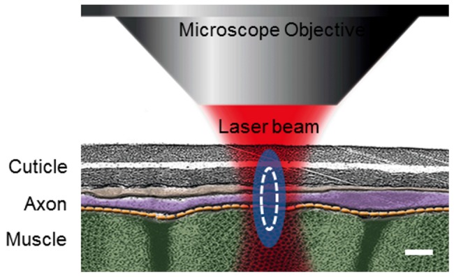
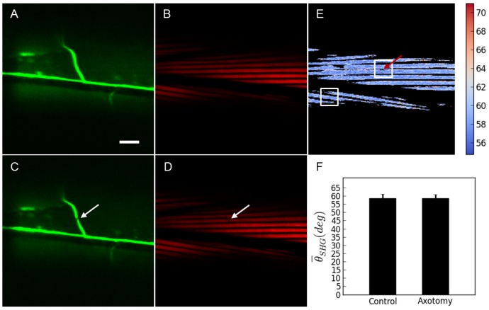
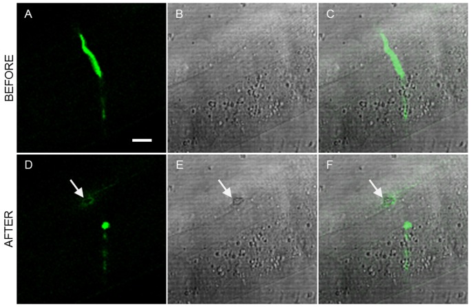
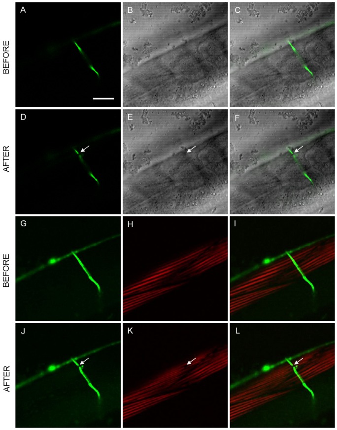
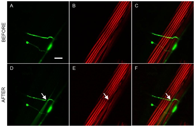
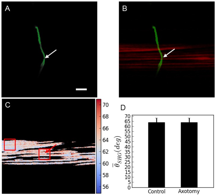
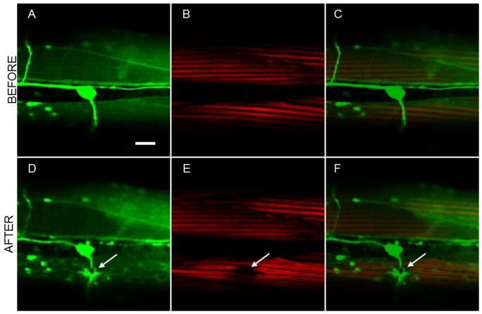
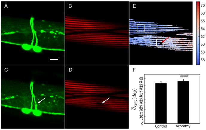
In this work highly localized femtosecond laser ablation is used to dissect single axons within a living Caenorhabditis elegans (C. elegans). We present a multimodal imaging methodology for the assessment of the collateral damage induced by the laser. This relies on the observation of the tissues surrounding the targeted region using a combination of different high resolution microscopy modalities. We present the use of Second Harmonic Generation (SHG) and Polarization Sensitive SHG (PSHG) to determine damage in the neighbor muscle cells. All the above is done using a single instrument: multimodal microscopy setup that allows simultaneous imaging in the linear and non-linear regimes and femtosecond-laser ablation.
在这项工作中,我们使用高度局域化的飞秒激光烧蚀技术来解剖活体秀丽隐杆线虫(C. elegans)中的单个轴突。我们提出了一种多模态成像方法来评估激光诱导的旁侧损伤。这依赖于使用不同高分辨率显微镜模式的组合来观察靶向区域周围的组织。我们提出使用二次谐波产生(SHG)和偏振敏感 SHG(PSHG)来确定邻近肌肉细胞的损伤。所有这些都是使用单个仪器完成的:多模态显微镜设置允许在线性和非线性状态以及飞秒激光烧蚀下同时成像。