• 文献检索
  • 文档翻译
  • 深度研究
  • 学术资讯
  • Suppr Zotero 插件Zotero 插件
  • 邀请有礼
  • 套餐&价格
  • 历史记录
应用&插件
Suppr Zotero 插件Zotero 插件浏览器插件Mac 客户端Windows 客户端微信小程序
定价
高级版会员购买积分包购买API积分包
服务
文献检索文档翻译深度研究API 文档MCP 服务
关于我们
关于 Suppr公司介绍联系我们用户协议隐私条款
关注我们

Suppr 超能文献

核心技术专利:CN118964589B侵权必究
粤ICP备2023148730 号-1Suppr @ 2026

文献检索

告别复杂PubMed语法,用中文像聊天一样搜索,搜遍4000万医学文献。AI智能推荐,让科研检索更轻松。

立即免费搜索

文件翻译

保留排版,准确专业,支持PDF/Word/PPT等文件格式,支持 12+语言互译。

免费翻译文档

深度研究

AI帮你快速写综述,25分钟生成高质量综述,智能提取关键信息,辅助科研写作。

立即免费体验

人与机器的较量:比较放射科医生对创伤性脑损伤患者脑部 MRI 的解读和 NeuroQuant®容积分析结果。

Man versus machine: comparison of radiologists' interpretations and NeuroQuant® volumetric analyses of brain MRIs in patients with traumatic brain injury.

机构信息

Virginia Institute of Neuropsychiatry, Midlothian, VA, USA.

出版信息

J Neuropsychiatry Clin Neurosci. 2013 Winter;25(1):32-9. doi: 10.1176/appi.neuropsych.11120377.

DOI:10.1176/appi.neuropsych.11120377
PMID:23487191
原文链接:https://pmc.ncbi.nlm.nih.gov/articles/PMC7185228/
Abstract

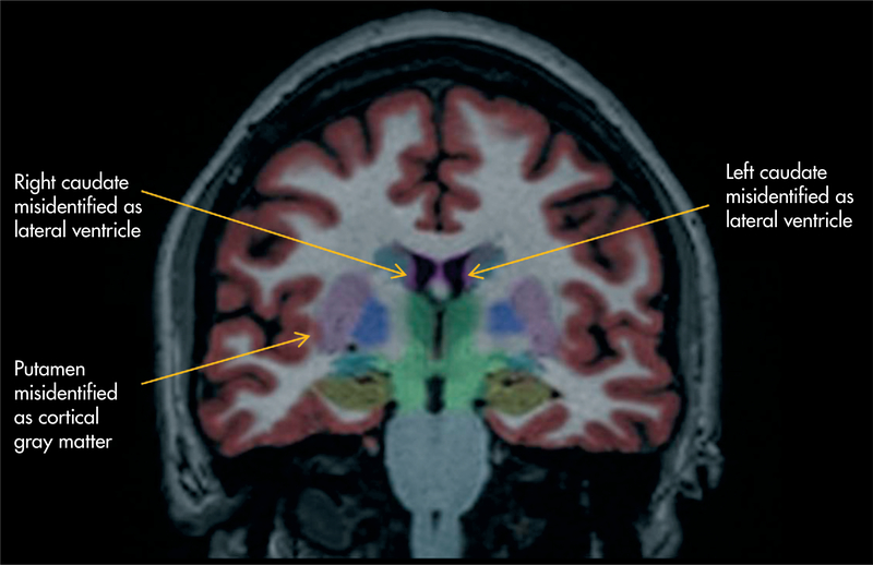
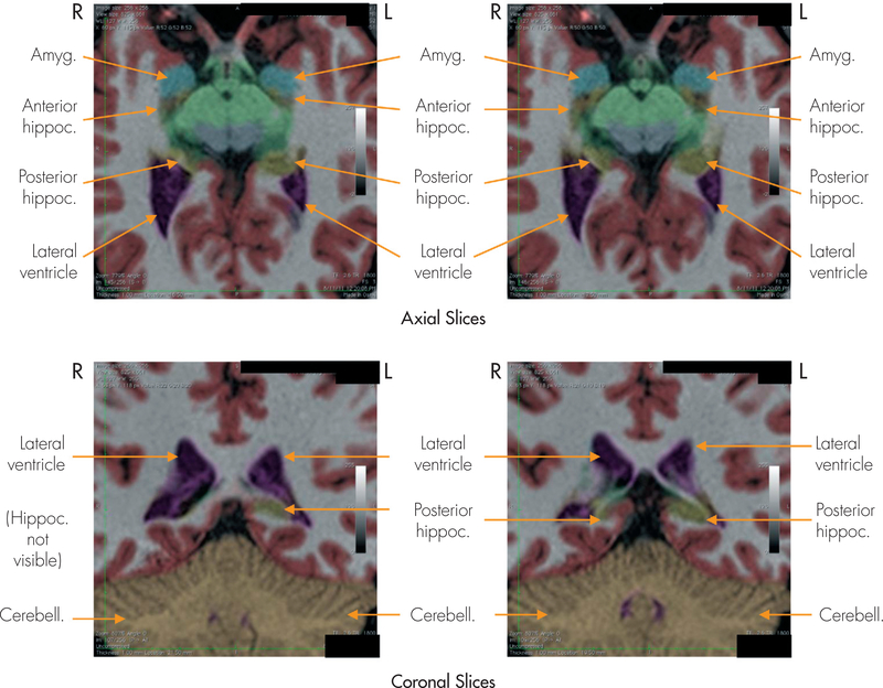
NeuroQuant® is a recently developed, FDA-approved software program for measuring brain MRI volume in clinical settings. The purpose of this study was to compare NeuroQuant with the radiologist's traditional approach, based on visual inspection, in 20 outpatients with mild or moderate traumatic brain injury (TBI). Each MRI was analyzed with NeuroQuant, and the resulting volumetric analyses were compared with the attending radiologist's interpretation. The radiologist's traditional approach found atrophy in 10.0% of patients; NeuroQuant found atrophy in 50.0% of patients. NeuroQuant was more sensitive for detecting brain atrophy than the traditional radiologist's approach.

摘要

NeuroQuant® 是一款最近开发的、获得 FDA 批准的软件程序,用于在临床环境下测量脑 MRI 容积。本研究的目的是在 20 名轻度或中度创伤性脑损伤(TBI)的门诊患者中,比较 NeuroQuant 与放射科医生基于视觉检查的传统方法。对每例 MRI 进行 NeuroQuant 分析,并将得出的容积分析与主治放射科医生的解读进行比较。放射科医生的传统方法发现 10.0%的患者有萎缩;NeuroQuant 发现 50.0%的患者有萎缩。NeuroQuant 比传统放射科医生的方法更敏感,能检测到脑萎缩。

https://cdn.ncbi.nlm.nih.gov/pmc/blobs/4253/7185228/d4d4c8cad67e/nihms-1557682-f0002.jpg
https://cdn.ncbi.nlm.nih.gov/pmc/blobs/4253/7185228/e674b82de829/nihms-1557682-f0001.jpg
https://cdn.ncbi.nlm.nih.gov/pmc/blobs/4253/7185228/d4d4c8cad67e/nihms-1557682-f0002.jpg
https://cdn.ncbi.nlm.nih.gov/pmc/blobs/4253/7185228/e674b82de829/nihms-1557682-f0001.jpg
https://cdn.ncbi.nlm.nih.gov/pmc/blobs/4253/7185228/d4d4c8cad67e/nihms-1557682-f0002.jpg

相似文献

1
Man versus machine: comparison of radiologists' interpretations and NeuroQuant® volumetric analyses of brain MRIs in patients with traumatic brain injury.人与机器的较量:比较放射科医生对创伤性脑损伤患者脑部 MRI 的解读和 NeuroQuant®容积分析结果。
J Neuropsychiatry Clin Neurosci. 2013 Winter;25(1):32-9. doi: 10.1176/appi.neuropsych.11120377.
2
Man Versus Machine Part 2: Comparison of Radiologists' Interpretations and NeuroQuant Measures of Brain Asymmetry and Progressive Atrophy in Patients With Traumatic Brain Injury.人与机器 第二部分:创伤性脑损伤患者中放射科医生的解读与NeuroQuant脑不对称和进行性萎缩测量的比较
J Neuropsychiatry Clin Neurosci. 2015;27(2):147-52. doi: 10.1176/appi.neuropsych.13040088.
3
NeuroQuant® and NeuroGage® reveal effects of traumatic brain injury on brain volume.NeuroQuant® 和 NeuroGage® 揭示了创伤性脑损伤对脑容量的影响。
Brain Inj. 2018;32(11):1437-1441. doi: 10.1080/02699052.2018.1489980. Epub 2018 Jun 28.
4
Comparison of Automated Brain Volume Measures obtained with NeuroQuant and FreeSurfer.使用NeuroQuant和FreeSurfer获得的脑容量自动测量结果的比较。
J Neuroimaging. 2015 Sep-Oct;25(5):721-7. doi: 10.1111/jon.12229. Epub 2015 Feb 26.
5
Updated Review of the Evidence Supporting the Medical and Legal Use of NeuroQuant and NeuroGage in Patients With Traumatic Brain Injury.支持NeuroQuant和NeuroGage在创伤性脑损伤患者中进行医学和法律应用的证据的更新综述。
Front Hum Neurosci. 2022 Apr 8;16:715807. doi: 10.3389/fnhum.2022.715807. eCollection 2022.
6
Progressive brain atrophy in patients with chronic neuropsychiatric symptoms after mild traumatic brain injury: a preliminary study.轻度创伤性脑损伤后慢性神经精神症状患者的进行性脑萎缩:一项初步研究。
Brain Inj. 2012;26(12):1500-9. doi: 10.3109/02699052.2012.694570. Epub 2012 Jun 21.
7
Hippocampal Sclerosis Detection with NeuroQuant Compared with Neuroradiologists.神经定量分析与神经放射科医师检测海马硬化的比较。
AJNR Am J Neuroradiol. 2020 Apr;41(4):591-597. doi: 10.3174/ajnr.A6454. Epub 2020 Mar 26.
8
Back to the future: estimating pre-injury brain volume in patients with traumatic brain injury.回到未来:估计创伤性脑损伤患者伤前脑容量
Neuroimage. 2014 Nov 15;102 Pt 2:565-78. doi: 10.1016/j.neuroimage.2014.07.043. Epub 2014 Aug 8.
9
Comparing Two Processing Pipelines to Measure Subcortical and Cortical Volumes in Patients with and without Mild Traumatic Brain Injury.比较两种处理流程以测量有和无轻度创伤性脑损伤患者的皮质下和皮质体积
J Neuroimaging. 2017 Jul;27(4):365-371. doi: 10.1111/jon.12431. Epub 2017 Feb 14.
10
Automated brain volumetrics in multiple sclerosis: a step closer to clinical application.多发性硬化症中的自动脑容量测量:向临床应用迈进了一步。
J Neurol Neurosurg Psychiatry. 2016 Jul;87(7):754-7. doi: 10.1136/jnnp-2015-312304. Epub 2016 Apr 12.

引用本文的文献

1
Association between BrainAGE and Alzheimer's disease biomarkers.脑龄(BrainAGE)与阿尔茨海默病生物标志物之间的关联。
Alzheimers Dement (Amst). 2025 Feb 27;17(1):e70094. doi: 10.1002/dad2.70094. eCollection 2025 Jan-Mar.
2
Neurofind: using deep learning to make individualised inferences in brain-based disorders.Neurofind:利用深度学习对脑部疾病进行个性化推断。
Transl Psychiatry. 2025 Feb 27;15(1):69. doi: 10.1038/s41398-025-03290-x.
3
Symptom Persistence Relates to Volume and Asymmetry of the Limbic System after Mild Traumatic Brain Injury.

本文引用的文献

1
NeuroQuant® revealed hippocampal atrophy in a patient with traumatic brain injury.NeuroQuant®检测显示一名创伤性脑损伤患者存在海马萎缩。
J Neuropsychiatry Clin Neurosci. 2012 Winter;24(1):E33. doi: 10.1176/appi.neuropsych.11020044.
2
Review of longitudinal studies of MRI brain volumetry in patients with traumatic brain injury.创伤性脑损伤患者脑MRI容积测量纵向研究综述
Brain Inj. 2011;25(13-14):1271-8. doi: 10.3109/02699052.2011.624568.
3
The Alzheimer's disease neuroimaging initiative: progress report and future plans.
轻度创伤性脑损伤后症状持续与边缘系统的体积和不对称性有关。
J Clin Med. 2024 Aug 30;13(17):5154. doi: 10.3390/jcm13175154.
4
Rate of abnormalities in quantitative MR neuroimaging of persons with chronic traumatic brain injury.慢性创伤性脑损伤患者定量磁共振神经影像学异常率。
BMC Neurol. 2024 Jul 5;24(1):235. doi: 10.1186/s12883-024-03745-6.
5
Structural neuroimaging markers of normal pressure hydrocephalus versus Alzheimer's dementia and Parkinson's disease, and hydrocephalus versus atrophy in chronic TBI-a narrative review.正常压力脑积水与阿尔茨海默病性痴呆和帕金森病的结构神经影像学标志物,以及慢性创伤性脑损伤中脑积水与萎缩的比较——一篇叙述性综述
Front Neurol. 2024 Mar 21;15:1347200. doi: 10.3389/fneur.2024.1347200. eCollection 2024.
6
Data-driven normative values based on generative manifold learning for quantitative MRI.基于生成流形学习的定量 MRI 数据驱动规范值。
Sci Rep. 2024 Mar 30;14(1):7563. doi: 10.1038/s41598-024-58141-4.
7
Forensic neurology: a distinct subspecialty at the intersection of neurology, neuroscience and law.法医神经病学:神经学、神经科学和法律交叉的独特亚专科。
Nat Rev Neurol. 2024 Mar;20(3):183-193. doi: 10.1038/s41582-023-00920-0. Epub 2024 Jan 16.
8
A real-world clinical validation for AI-based MRI monitoring in multiple sclerosis.基于人工智能的磁共振成像监测在多发性硬化症中的真实世界临床验证。
NPJ Digit Med. 2023 Oct 19;6(1):196. doi: 10.1038/s41746-023-00940-6.
9
Journey to the other side of the brain: asymmetry in patients with chronic mild or moderate traumatic brain injury.大脑另一侧之旅:慢性轻度或中度创伤性脑损伤患者的不对称性
Concussion. 2023 Jan 31;8(1):CNC101. doi: 10.2217/cnc-2022-0003. eCollection 2022 Dec.
10
Cortical Grey matter volume depletion links to neurological sequelae in post COVID-19 "long haulers".皮质灰质体积减少与新冠后“长新冠”患者的神经后遗症相关。
BMC Neurol. 2023 Jan 17;23(1):22. doi: 10.1186/s12883-023-03049-1.
阿尔茨海默病神经影像学倡议:进展报告和未来计划。
Alzheimers Dement. 2010 May;6(3):202-11.e7. doi: 10.1016/j.jalz.2010.03.007.
4
Trajectories of brain aging in middle-aged and older adults: regional and individual differences.中年人及老年人的大脑老化轨迹:区域和个体差异。
Neuroimage. 2010 Jun;51(2):501-11. doi: 10.1016/j.neuroimage.2010.03.020. Epub 2010 Mar 16.
5
Alzheimer's Disease Neuroimaging Initiative (ADNI): clinical characterization.阿尔茨海默病神经影像学倡议(ADNI):临床特征。
Neurology. 2010 Jan 19;74(3):201-9. doi: 10.1212/WNL.0b013e3181cb3e25. Epub 2009 Dec 30.
6
Intra- and interscanner variability of automated voxel-based volumetry based on a 3D probabilistic atlas of human cerebral structures.基于人体脑结构三维概率图谱的自动体素容积测量的扫描仪内和扫描仪间可变性。
Neuroimage. 2010 Feb 1;49(3):2216-24. doi: 10.1016/j.neuroimage.2009.10.066. Epub 2009 Oct 28.
7
Fully-automated volumetric MRI with normative ranges: translation to clinical practice.具有正常范围的全自动容积磁共振成像:向临床实践的转化
Behav Neurol. 2009;21(1):21-8. doi: 10.3233/BEN-2009-0226.
8
High-throughput, fully automated volumetry for prediction of MMSE and CDR decline in mild cognitive impairment.用于预测轻度认知障碍中简易精神状态检查表(MMSE)和临床痴呆评定量表(CDR)下降的高通量、全自动容积测定法。
Alzheimer Dis Assoc Disord. 2009 Apr-Jun;23(2):139-45. doi: 10.1097/WAD.0b013e318192e745.
9
Fully-automated quantification of regional brain volumes for improved detection of focal atrophy in Alzheimer disease.全自动定量分析区域脑容量以改善阿尔茨海默病中局灶性萎缩的检测。
AJNR Am J Neuroradiol. 2009 Mar;30(3):578-80. doi: 10.3174/ajnr.A1402. Epub 2008 Dec 26.
10
The Alzheimer's Disease Neuroimaging Initiative (ADNI): MRI methods.阿尔茨海默病神经影像学倡议(ADNI):磁共振成像方法
J Magn Reson Imaging. 2008 Apr;27(4):685-91. doi: 10.1002/jmri.21049.