1. Department of Endodontic, Dental Research Center of Shahid Beheshti University of Medical Science, Tehran, Iran.
Cell J. 2012 Winter;13(4):223-8. Epub 2011 Dec 22.
Evaluation of the effect of Propolis as a bioactive material on quality of dentin and presence of dental pulp stem cells.
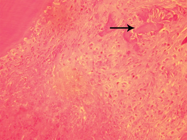
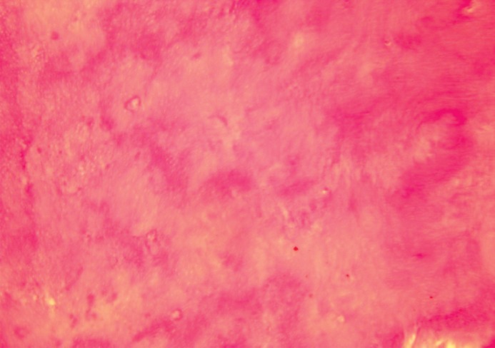
For conducting this experimental split-mouth study,a total of 48 maxillary and mandibular incisors of male guinea pigs were randomly divided into an experimental Propolis group and a control calcium hydroxide group. Cutting the crowns and using Propolis or calcium hydroxide to cap the pulp, all of the cavities were sealed. Sections of the teeth were obtained after sacrificing 4 guinea pigs from each group on the 10(th), 15(th) and 30(th) day. After they had been stained by hematoxylin and eosin (H&E), specimens underwent a histological evaluation under a light microscope for identification of the presence of odontoblast-like cells, pulp vitality, congestion, inflammation of the pulp and the presence of remnants of the material used. The immunohistochemistry (IHC) method using CD29 and CD146 was performed to evaluate the presence of stem cells and the results were statistically evaluated by Kruskal-Wallis, Chi Square and Fisher tests.
In H&E stained specimens, there was no difference between the two groups in the presence of odontoblast-like cells, pulp vitality, congestion, inflammation of the pulp and the presence of remnants of used material(p>0.05). There was a significant difference between the quality of regenerative dentin on the 15(th) and 30(th) days (p<0.05): all of the Propolis cases presented tubular dentin while 14% of the calcium hydroxide cases produced porous dentin. There was no significant difference between Propolis and calcium hydroxide in stimulation of dental pulp stem cells (DPSCs).
This study which is the first one that documented the stimulation of stem cells by Propolis, provides evidence that this material has advantages over calcium hydroxide as a capping agent in vital pulp therapy. In addition to producing no pulpal inflammation, infection or necrosis this material induces the production of high quality tubular dentin.
评价蜂胶作为一种生物活性物质对牙本质质量和牙髓干细胞存在的影响。
为进行这项实验性的分口研究,将 48 颗雄性豚鼠的上颌和下颌切牙随机分为蜂胶实验组和对照组的氢氧化钙组。切冠后用蜂胶或氢氧化钙盖髓,所有窝洞均密封。每组处死 4 只豚鼠后,于第 10、15、30 天分别获得牙齿的切片。经过苏木精-伊红(H&E)染色后,在光镜下对标本进行组织学评价,以鉴定有无成牙本质细胞样细胞、牙髓活力、充血、炎症和所用材料的残余物。采用 CD29 和 CD146 免疫组织化学(IHC)方法评价干细胞的存在,并采用 Kruskal-Wallis、卡方和 Fisher 检验对结果进行统计学评价。
在 H&E 染色标本中,两组间成牙本质细胞样细胞的存在、牙髓活力、充血、炎症和所用材料残余物的存在无差异(p>0.05)。在第 15 和 30 天,再生牙本质的质量有显著差异(p<0.05):所有蜂胶组均表现为管状牙本质,而 14%的氢氧化钙组产生多孔牙本质。蜂胶和氢氧化钙对牙髓干细胞(DPSCs)的刺激无显著差异。
这项研究首次记录了蜂胶对干细胞的刺激作用,证明了这种材料在活髓治疗中作为盖髓剂优于氢氧化钙。除了不引起牙髓炎症、感染或坏死外,这种材料还诱导产生高质量的管状牙本质。