Asgary Saeed, Parirokh Masoud, Eghbal Mohammad Jafar, Ghoddusi Jamileh
Department of Endodontics, Iranian Center for Endodontic Research, Dental School, Shaheed Beheshti University of Medical Sciences, Tehran, Iran.
Department of Endodontics, Dental School, Kerman University of Medical Sciences, Kerman, Iran.
Iran Endod J. 2006 Winter;1(4):117-23. Epub 2007 Jan 20.
This investigation evaluates the effects of mineral trioxide aggregate (MTA), calcium hydroxide (CH) and calcium enriched mixture (CEM) as pulp capping materials on dental pulp tissues.
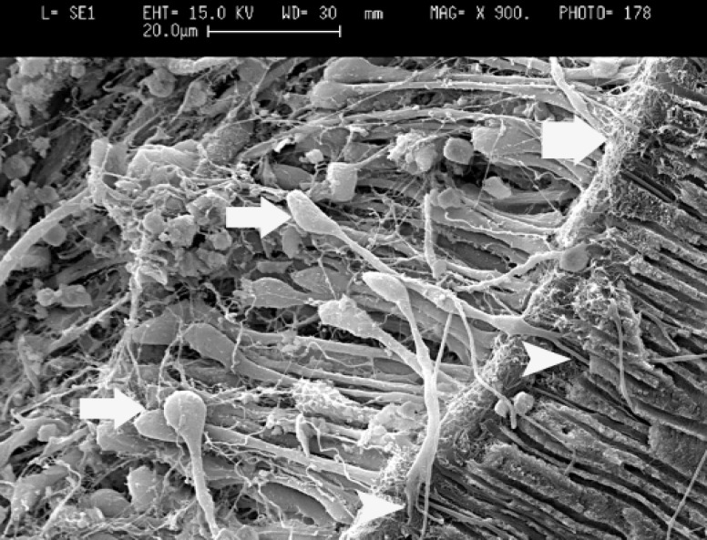
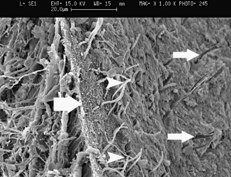
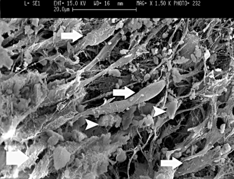
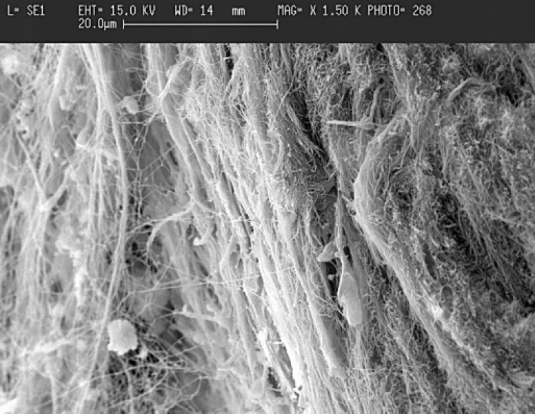
The experimental procedures were performed on eighteen intact dog canine teeth. The pulps were exposed. Cavities were randomly filled with CEM, MTA, or CH followed by glass ionomer filling. After 2 months, animals were sacrificed, each tooth was sectioned into halves, and the interface between each capping material and pulp tissue was evaluated by scanning electron microscope (SEM) in profile view of the specimens.
Dentinal bridge formation as the most characteristic reaction was resulted from SEM observation in all examined groups. Odontoblast-like cells were formed and create dens collagen network, which was calcified gradually by deposition of calcosphirit structures to form newly dentinal bridge.
Based on the results of this in vivo study, it was concluded that these test materials are able to produce calcified tissue in underlying pulp in the case of being used as a pulp capping agent. Additionally, it appears that CEM has the potential to be used as a direct pulp capping material during vital pulp therapy.
本研究评估了三氧化矿物凝聚体(MTA)、氢氧化钙(CH)和富钙混合物(CEM)作为盖髓材料对牙髓组织的影响。
在18颗完整的犬尖牙上进行实验操作。暴露牙髓。窝洞随机用CEM、MTA或CH填充,然后用玻璃离子充填。2个月后,处死动物,将每颗牙齿切成两半,通过扫描电子显微镜(SEM)在标本的侧面视图中评估每种盖髓材料与牙髓组织之间的界面。
扫描电镜观察显示,所有检测组均出现了作为最典型反应的牙本质桥形成。形成了成牙本质细胞样细胞并形成致密的胶原网络,该网络通过钙球晶结构的沉积逐渐钙化,形成新的牙本质桥。
基于本体内研究结果,得出结论:这些测试材料用作盖髓剂时能够在下方牙髓中产生钙化组织。此外,似乎CEM有潜力在活髓治疗期间用作直接盖髓材料。