Ingo Carson, Magin Richard L, Colon-Perez Luis, Triplett William, Mareci Thomas H
Department of Bioengineering, University of Illinois at Chicago, Chicago, Illinois, USA.
Magn Reson Med. 2014 Feb;71(2):617-27. doi: 10.1002/mrm.24706.
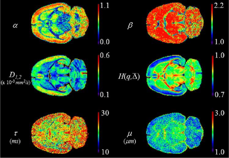
In diffusion-weighted MRI studies of neural tissue, the classical model assumes the statistical mechanics of Brownian motion and predicts a monoexponential signal decay. However, there have been numerous reports of signal decays that are not monoexponential, particularly in the white matter.
We modeled diffusion in neural tissue from the perspective of the continuous time random walk. The characteristic diffusion decay is represented by the Mittag-Leffler function, which relaxes a priori assumptions about the governing statistics. We then used entropy as a measure of the anomalous features for the characteristic function.
Diffusion-weighted MRI experiments were performed on a fixed rat brain using an imaging spectrometer at 17.6 T with b-values arrayed up to 25,000 s/mm(2). Additionally, we examined the impact of varying either the gradient strength, q, or mixing time, Δ, on the observed diffusion dynamics.
In white and gray matter regions, the Mittag-Leffler and entropy parameters demonstrated new information regarding subdiffusion and produced different image contrast from that of the classical diffusion coefficient. The choice of weighting on q and Δ produced different image contrast within the regions of interest.
We propose these parameters have the potential as biomarkers for morphology in neural tissue.
在神经组织的扩散加权磁共振成像(MRI)研究中,经典模型假定布朗运动的统计力学,并预测单指数信号衰减。然而,已有大量关于非单指数信号衰减的报道,尤其是在白质中。
我们从连续时间随机游走的角度对神经组织中的扩散进行建模。特征扩散衰减由米塔格 - 莱夫勒函数表示,该函数放宽了关于主导统计量的先验假设。然后,我们使用熵作为特征函数异常特征的度量。
使用成像光谱仪在17.6 T磁场下对固定的大鼠脑进行扩散加权MRI实验,b值高达25,000 s/mm²。此外,我们研究了改变梯度强度q或混合时间Δ对观察到的扩散动力学的影响。
在白质和灰质区域,米塔格 - 莱夫勒和熵参数展示了关于亚扩散的新信息,并产生了与经典扩散系数不同的图像对比度。对q和Δ的加权选择在感兴趣区域内产生了不同的图像对比度。
我们认为这些参数有潜力作为神经组织形态学的生物标志物。