• 文献检索
  • 文档翻译
  • 深度研究
  • 学术资讯
  • Suppr Zotero 插件Zotero 插件
  • 邀请有礼
  • 套餐&价格
  • 历史记录
应用&插件
Suppr Zotero 插件Zotero 插件浏览器插件Mac 客户端Windows 客户端微信小程序
定价
高级版会员购买积分包购买API积分包
服务
文献检索文档翻译深度研究API 文档MCP 服务
关于我们
关于 Suppr公司介绍联系我们用户协议隐私条款
关注我们

Suppr 超能文献

核心技术专利:CN118964589B侵权必究
粤ICP备2023148730 号-1Suppr @ 2026

文献检索

告别复杂PubMed语法,用中文像聊天一样搜索,搜遍4000万医学文献。AI智能推荐,让科研检索更轻松。

立即免费搜索

文件翻译

保留排版,准确专业,支持PDF/Word/PPT等文件格式,支持 12+语言互译。

免费翻译文档

深度研究

AI帮你快速写综述,25分钟生成高质量综述,智能提取关键信息,辅助科研写作。

立即免费体验

IMP-3 在角化棘皮瘤和皮肤鳞状细胞癌中的表达:免疫组织化学研究。

IMP-3 expression in keratoacanthomas and squamous cell carcinomas of the skin: an immunohistochemical study.

机构信息

Department of Surgical Sciences, Division of Pathology, University of Cagliari, Italy.

出版信息

Eur J Histochem. 2013 Feb 14;57(1):e6. doi: 10.4081/ejh.2013.e6.

DOI:10.4081/ejh.2013.e6
PMID:23549465
原文链接:https://pmc.ncbi.nlm.nih.gov/articles/PMC3683613/
Abstract

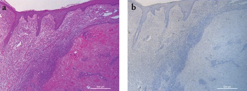
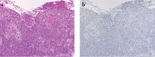
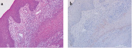
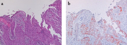
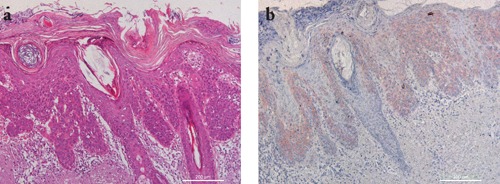
The protein insulin-like growth factor II mRNA binding protein 3 (IMP3) is an important factor for cell migration and adhesion in malignancies. Recent studies have shown a remarkable overexpression of IMP3 in different human malignant neoplasms and also revealed it as an important prognostic marker in some tumor entities. The purpose of this study is to compare IMP3 immunostaining in squamous cellular skin tumor and determine whether IMP3 can aid in the differential diagnosis of these lesions. To our knowledge, IMP3 expression has not been investigated in skin squamous cell proliferations thus far. Immunohistochemical staining for IMP3 was performed on slides organized by samples from 67 patients, 34 with keratoacanthoma and 33 with primary squamous cell carcinoma (16 invasive and 17 in situ). The majority of our KAs (25/34) were negative for IMP-3 staining. The majority of SCCs (19/33) are positive for IMP3 staining. The percentage of IMP3 positive cells increases significantly in group SCC (p=0.0111), and in particular in the SCC in situ group (p=0.0021) with respect to the KA group.  IMP3 intensity staining increases significantly in SCCs (p=0.0213), and particularly in SCCs (p=0.008) with respect to KA. Our data show that IMP3 expression is different in keratoacanthomas with respect to squamous cell carcinoma. IMP3 assessment and staining pattern, together with a careful histological study, can be useful in the differential diagnosis between KA e SCC.

摘要

胰岛素样生长因子 II mRNA 结合蛋白 3(IMP3)是恶性肿瘤中细胞迁移和黏附的重要因素。最近的研究表明,IMP3 在不同的人类恶性肿瘤中显著过表达,并且在一些肿瘤实体中也被揭示为重要的预后标志物。本研究的目的是比较鳞状细胞皮肤肿瘤中的 IMP3 免疫染色,并确定 IMP3 是否有助于这些病变的鉴别诊断。据我们所知,迄今为止,IMP3 在皮肤鳞状细胞增生中的表达尚未被研究过。对 67 名患者的样本进行了 IMP3 免疫组织化学染色,其中 34 名患者为角化棘皮瘤,33 名患者为原发性鳞状细胞癌(16 名侵袭性和 17 名原位)。我们的大多数角化棘皮瘤(25/34)IMP-3 染色为阴性。大多数鳞状细胞癌(19/33)IMP3 染色阳性。与 KA 组相比,SCC 组(p=0.0111),特别是 SCC 原位组(p=0.0021)IMP3 阳性细胞的百分比明显增加。与 KA 相比,SCCs(p=0.0213)和尤其是 SCCs(p=0.008)中 IMP3 强度染色明显增加。我们的数据表明,IMP3 在角化棘皮瘤与鳞状细胞癌中的表达不同。IMP3 评估和染色模式,结合仔细的组织学研究,可用于 KA 和 SCC 之间的鉴别诊断。

https://cdn.ncbi.nlm.nih.gov/pmc/blobs/c097/3683613/2e39131469a8/ejh-2013-1-e6-g005.jpg
https://cdn.ncbi.nlm.nih.gov/pmc/blobs/c097/3683613/da72fda05085/ejh-2013-1-e6-g001.jpg
https://cdn.ncbi.nlm.nih.gov/pmc/blobs/c097/3683613/c53e4315bcbe/ejh-2013-1-e6-g002.jpg
https://cdn.ncbi.nlm.nih.gov/pmc/blobs/c097/3683613/bcae62f1a173/ejh-2013-1-e6-g003.jpg
https://cdn.ncbi.nlm.nih.gov/pmc/blobs/c097/3683613/e432cd6ff06d/ejh-2013-1-e6-g004.jpg
https://cdn.ncbi.nlm.nih.gov/pmc/blobs/c097/3683613/2e39131469a8/ejh-2013-1-e6-g005.jpg

相似文献

1
IMP-3 expression in keratoacanthomas and squamous cell carcinomas of the skin: an immunohistochemical study.IMP-3 在角化棘皮瘤和皮肤鳞状细胞癌中的表达:免疫组织化学研究。
Eur J Histochem. 2013 Feb 14;57(1):e6. doi: 10.4081/ejh.2013.e6.
2
Insulin-like growth factor 2 mRNA-binding protein-3 as a marker for distinguishing between cutaneous squamous cell carcinoma and keratoacanthoma.胰岛素样生长因子2信使核糖核酸结合蛋白-3作为区分皮肤鳞状细胞癌和角化棘皮瘤的标志物
Int J Oncol. 2016 Mar;48(3):1007-15. doi: 10.3892/ijo.2016.3323. Epub 2016 Jan 5.
3
p16 expression in keratoacanthomas and squamous cell carcinomas of the skin: an immunohistochemical study.皮肤角化棘皮瘤和鳞状细胞癌中p16的表达:一项免疫组织化学研究
Arch Pathol Lab Med. 2006 Jan;130(1):69-73. doi: 10.5858/2006-130-69-PEIKAS.
4
Immunohistochemical staining for desmogleins 1 and 2 in keratinocytic neoplasms with squamous phenotype: actinic keratosis, keratoacanthoma and squamous cell carcinoma of the skin.具有鳞状表型的角质形成细胞肿瘤(光化性角化病、角化棘皮瘤和皮肤鳞状细胞癌)中桥粒芯糖蛋白1和2的免疫组织化学染色
Br J Cancer. 1998 Apr;77(8):1275-9. doi: 10.1038/bjc.1998.213.
5
[The value of laminin-322 staining in distinguishing between keratoacanthoma, keratoacanthoma with areas of squamous cell carcinoma, and crateriform squamous cell carcinoma].[层粘连蛋白-322染色在鉴别角化棘皮瘤、伴有鳞状细胞癌区域的角化棘皮瘤和火山口状鳞状细胞癌中的价值]
Actas Dermosifiliogr. 2012 May;103(4):308-16. doi: 10.1016/j.ad.2011.09.004. Epub 2011 Dec 15.
6
The level of syndecan-1 expression is a distinguishing feature in behavior between keratoacanthoma and invasive cutaneous squamous cell carcinoma.Syndecan-1的表达水平是角化棘皮瘤和侵袭性皮肤鳞状细胞癌行为的一个显著特征。
Mod Pathol. 2002 Jan;15(1):45-9. doi: 10.1038/modpathol.3880488.
7
Beta-catenin and CD44 expression in keratoacanthoma and squamous cell carcinoma of the skin.
Tumori. 2007 May-Jun;93(3):284-9. doi: 10.1177/030089160709300310.
8
Stromal expression of CD34, α-smooth muscle actin and CD26/DPPIV in squamous cell carcinoma of the skin: a comparative immunohistochemical study.皮肤鳞状细胞癌中 CD34、α-平滑肌肌动蛋白和 CD26/DPPIV 的基质表达:一项比较免疫组织化学研究。
Pathol Oncol Res. 2012 Jan;18(1):25-31. doi: 10.1007/s12253-011-9412-9. Epub 2011 Jun 15.
9
Keratoacanthoma versus squamous cell carcinoma. An immunohistochemical reappraisal of p53 protein and proliferating cell nuclear antigen expression in keratoacanthoma-like tumors.角化棘皮瘤与鳞状细胞癌。对角化棘皮瘤样肿瘤中p53蛋白和增殖细胞核抗原表达的免疫组织化学重新评估。
Am J Dermatopathol. 1995 Aug;17(4):324-31. doi: 10.1097/00000372-199508000-00003.
10
IMP3 is a novel biomarker to predict metastasis and prognosis of tongue squamous cell carcinoma.IMP3是一种预测舌鳞状细胞癌转移和预后的新型生物标志物。
J Craniofac Surg. 2011 Nov;22(6):2022-5. doi: 10.1097/SCS.0b013e3182319750.

引用本文的文献

1
Identifying SCC Lesions Capable of Spontaneous Regression by Using Immunohistochemistry: A Systematic Review and Meta-Analysis.利用免疫组织化学鉴定能够自发消退的鳞状细胞癌病变:一项系统评价和荟萃分析
Dermatol Pract Concept. 2024 Apr 1;14(2):e2024047. doi: 10.5826/dpc.1402a47.
2
IMP3 Expression as a Potential Tumour Marker in High-Risk Localisations of Cutaneous Squamous Cell Carcinoma: IMP3 in Metastatic cSCC.IMP3表达作为皮肤鳞状细胞癌高危部位潜在的肿瘤标志物:转移性皮肤鳞状细胞癌中的IMP3
Cancers (Basel). 2023 Aug 14;15(16):4087. doi: 10.3390/cancers15164087.
3
The Contributions of Cancer-Testis and Developmental Genes to the Pathogenesis of Keratinocyte Carcinomas.

本文引用的文献

1
Insulin-like growth factor 2 messenger RNA binding protein 3 (IGF2BP3) is a marker of unfavourable prognosis in colorectal cancer.胰岛素样生长因子 2 信使 RNA 结合蛋白 3(IGF2BP3)是结直肠癌预后不良的标志物。
Eur J Cancer. 2012 Dec;48(18):3405-13. doi: 10.1016/j.ejca.2012.06.021. Epub 2012 Jul 26.
2
Expression of insulin-like growth factor II mRNA-binding protein 3 in squamous cell carcinomas of the head and neck.胰岛素样生长因子 II mRNA 结合蛋白 3 在头颈部鳞状细胞癌中的表达。
J Oral Pathol Med. 2013 Feb;42(2):125-32. doi: 10.1111/j.1600-0714.2012.01178.x. Epub 2012 May 29.
3
The usefulness of c-Kit in the immunohistochemical assessment of melanocytic lesions.
癌-睾丸基因和发育基因对角化细胞癌发病机制的作用
Cancers (Basel). 2022 Jul 26;14(15):3630. doi: 10.3390/cancers14153630.
4
Diagnostic Utility of Immunohistochemical Expressions of IMP3 Versus DOG1 and p63 in Salivary Gland Tumors.免疫组织化学表达 IMP3 与 DOG1 和 p63 在唾液腺肿瘤中的诊断效用。
Turk Patoloji Derg. 2020;36(3):227-236. doi: 10.5146/tjpath.2020.01496.
5
Exploration of potential key pathways and genes in multiple ocular cancers through bioinformatics analysis.通过生物信息学分析探索多种眼部癌症中的潜在关键通路和基因。
Graefes Arch Clin Exp Ophthalmol. 2019 Oct;257(10):2329-2341. doi: 10.1007/s00417-019-04410-2. Epub 2019 Jul 15.
6
IMP3 overexpression occurs in various important cancer types and is linked to aggressive tumor features: A tissue microarray study on 8,877 human cancers and normal tissues.IMP3 过表达发生在各种重要的癌症类型中,并与侵袭性肿瘤特征相关:在 8877 个人类癌症和正常组织的组织微阵列研究中。
Oncol Rep. 2018 Jan;39(1):3-12. doi: 10.3892/or.2017.6072. Epub 2017 Nov 2.
7
IGF2BP3 as a potential tissue marker for the diagnosis of esophageal high-grade intraepithelial neoplasia.胰岛素样生长因子2 mRNA结合蛋白3作为诊断食管高级别上皮内瘤变的潜在组织标志物
Onco Targets Ther. 2017 Aug 1;10:3861-3866. doi: 10.2147/OTT.S141179. eCollection 2017.
8
IMP3 expression in small-intestine neuroendocrine neoplasms: a new predictor of recurrence.IMP3 在小肠神经内分泌肿瘤中的表达:复发的新预测因子。
Endocrine. 2017 Nov;58(2):360-367. doi: 10.1007/s12020-017-1249-x. Epub 2017 Feb 17.
9
Insulin-like growth factor 2 mRNA-binding protein-3 as a marker for distinguishing between cutaneous squamous cell carcinoma and keratoacanthoma.胰岛素样生长因子2信使核糖核酸结合蛋白-3作为区分皮肤鳞状细胞癌和角化棘皮瘤的标志物
Int J Oncol. 2016 Mar;48(3):1007-15. doi: 10.3892/ijo.2016.3323. Epub 2016 Jan 5.
10
Histochemistry in biology and medicine: a message from the citing journals.生物学与医学中的组织化学:来自引用期刊的信息。
Eur J Histochem. 2015 Dec 23;59(4):2610. doi: 10.4081/ejh.2015.2610.
c-Kit 在黑色素细胞病变免疫组织化学评估中的作用。
Eur J Histochem. 2011 Jun 7;55(2):e20. doi: 10.4081/ejh.2011.e20.
4
IMP3 is a novel biomarker to predict metastasis and prognosis of gastric adenocarcinoma: a retrospective study.IMP3 是一种预测胃腺癌转移和预后的新型生物标志物:一项回顾性研究。
Chin Med J (Engl). 2010 Dec;123(24):3554-8.
5
IMP3 is a novel biomarker to predict metastasis and prognosis of tongue squamous cell carcinoma.IMP3是一种预测舌鳞状细胞癌转移和预后的新型生物标志物。
J Craniofac Surg. 2011 Nov;22(6):2022-5. doi: 10.1097/SCS.0b013e3182319750.
6
IMP3, a new biomarker to predict progression of cervical intraepithelial neoplasia into invasive cancer.IMP3,一种预测宫颈上皮内瘤变进展为浸润性癌的新型生物标志物。
Am J Surg Pathol. 2011 Nov;35(11):1638-45. doi: 10.1097/PAS.0b013e31823272d4.
7
Diagnostic value of immunohistochemical IMP3 expression in core needle biopsies of pancreatic ductal adenocarcinoma.免疫组织化学 IMP3 表达在胰腺导管腺癌核心针活检中的诊断价值。
Am J Surg Pathol. 2011 Jun;35(6):873-7. doi: 10.1097/PAS.0b013e3182189223.
8
IMP3 expression in urothelial carcinomas of the urinary bladder.IMP3在膀胱尿路上皮癌中的表达。
Turk Patoloji Derg. 2011 Jan;27(1):31-7. doi: 10.5146/tjpath.2010.01044.
9
IMP3 distinguishes uterine serous carcinoma from endometrial endometrioid adenocarcinoma.IMP3 可区分子宫浆液性癌和子宫内膜样腺癌。
Am J Clin Pathol. 2010 Jun;133(6):899-908. doi: 10.1309/AJCPQDQXJ4FNRFQB.
10
Squamous cell carcinoma arising in keratoacanthoma: a neglected phenomenon in the elderly.角化棘皮瘤中发生的鳞状细胞癌:老年人中一个被忽视的现象。
Am J Dermatopathol. 2010 Jul;32(5):423-6. doi: 10.1097/DAD.0b013e3181c4340a.