• 文献检索
  • 文档翻译
  • 深度研究
  • 学术资讯
  • Suppr Zotero 插件Zotero 插件
  • 邀请有礼
  • 套餐&价格
  • 历史记录
应用&插件
Suppr Zotero 插件Zotero 插件浏览器插件Mac 客户端Windows 客户端微信小程序
定价
高级版会员购买积分包购买API积分包
服务
文献检索文档翻译深度研究API 文档MCP 服务
关于我们
关于 Suppr公司介绍联系我们用户协议隐私条款
关注我们

Suppr 超能文献

核心技术专利:CN118964589B侵权必究
粤ICP备2023148730 号-1Suppr @ 2026

文献检索

告别复杂PubMed语法,用中文像聊天一样搜索,搜遍4000万医学文献。AI智能推荐,让科研检索更轻松。

立即免费搜索

文件翻译

保留排版,准确专业,支持PDF/Word/PPT等文件格式,支持 12+语言互译。

免费翻译文档

深度研究

AI帮你快速写综述,25分钟生成高质量综述,智能提取关键信息,辅助科研写作。

立即免费体验

破骨细胞吸收表面的独特亚区。

Distinctive subdomains in the resorbing surface of osteoclasts.

机构信息

Division of Basic Medical Sciences, St George's, University of London, Cranmer Terrace, Tooting, London, United Kingdom.

出版信息

PLoS One. 2013;8(3):e60285. doi: 10.1371/journal.pone.0060285. Epub 2013 Mar 21.

DOI:10.1371/journal.pone.0060285
PMID:23555944
原文链接:https://pmc.ncbi.nlm.nih.gov/articles/PMC3605329/
Abstract

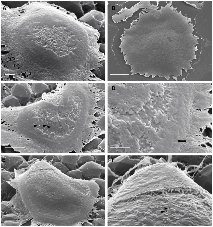
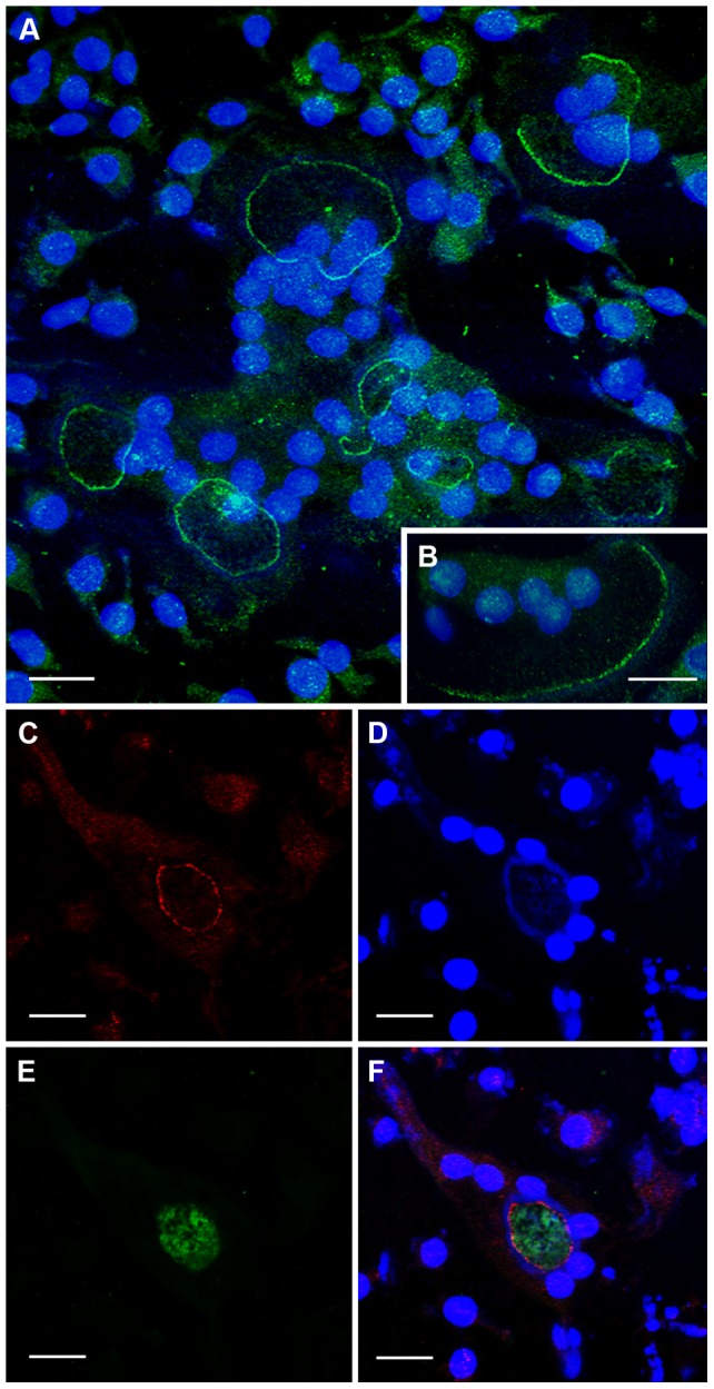
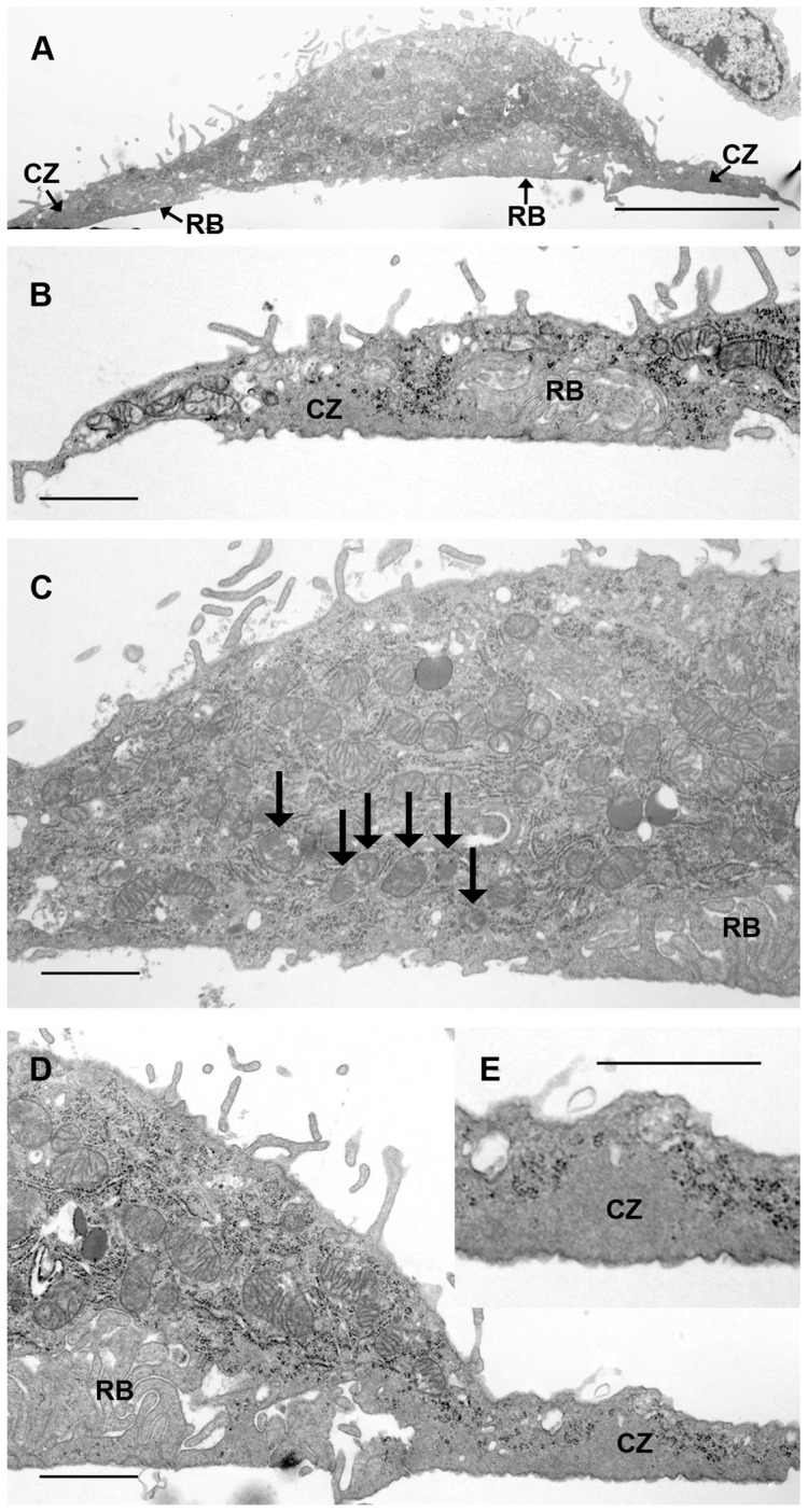
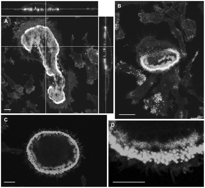
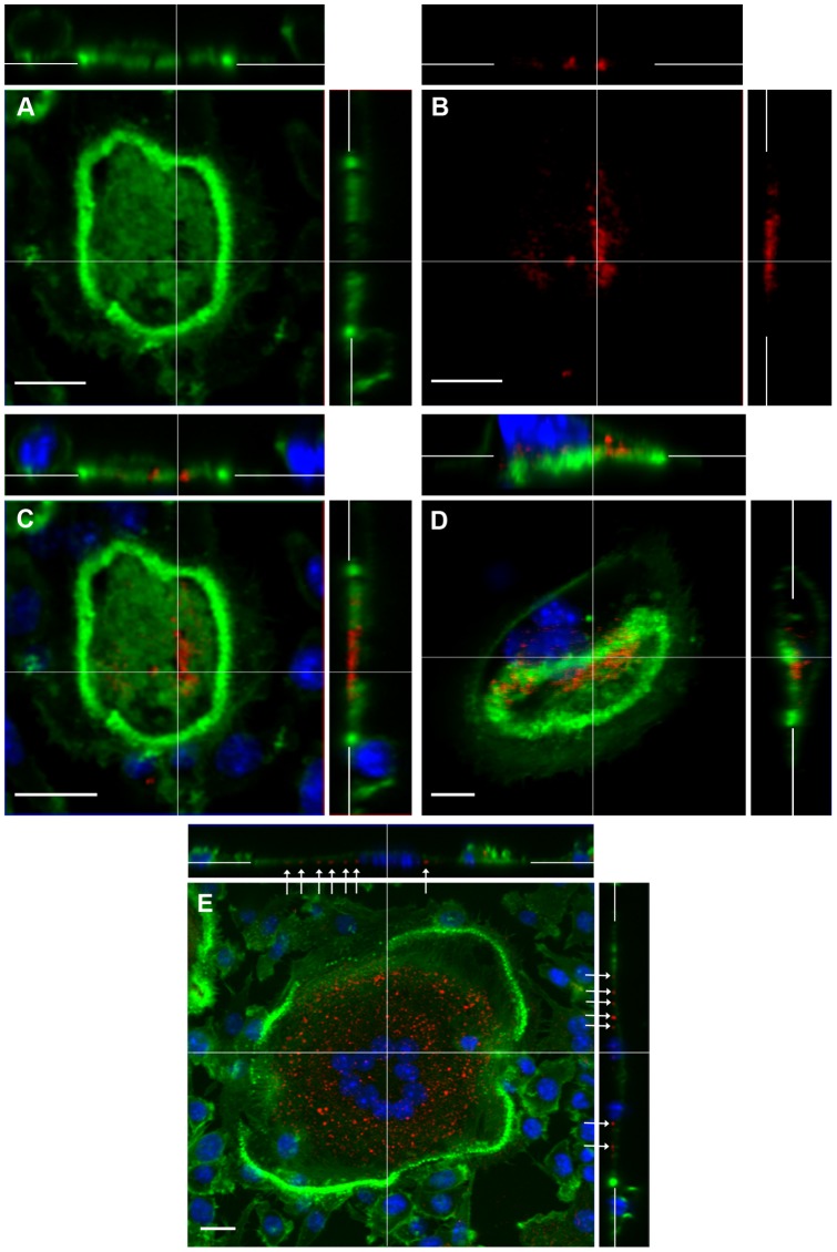
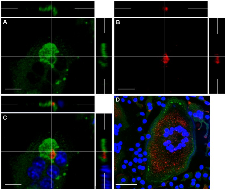
We employed a novel technique to inspect the substrate-apposed surface of activated osteoclasts, the cells that resorb bone, in the scanning electron microscope. The surface revealed unexpected complexity. At the periphery of the cells were circles and crescents of individual or confluent nodules. These corresponded to the podosomes and actin rings that form a 'sealing zone', encircling the resorptive hemivacuole into which protons and enzymes are secreted. Inside these rings and crescents the osteoclast surface was covered with strips and patches of membrane folds, which were flattened against the substrate surface and surrounded by fold-free membrane in which many orifices could be seen. Corresponding regions of folded and fold-free membrane were found by transmission electron microscopy in osteoclasts incubated on bone. We correlated these patterns with the distribution of several proteins crucial to resorption. The strips and patches of membrane folds corresponded in distribution to vacuolar H+-ATPase, and frequently co-localized with F-actin. Cathepsin K localized to F-actin-free foci towards the center of cells with circular actin rings, and at the retreating pole of cells with actin crescents. The chloride/proton antiporter ClC-7 formed a sharply-defined band immediately inside the actin ring, peripheral to vacuolar H+-ATPase. The sealing zone of osteoclasts is permeable to molecules with molecular mass up to 10,000. Therefore, ClC-7 might be distributed at the periphery of the resorptive hemivacuole in order to prevent protons from escaping laterally from the hemivacuole into the sealing zone, where they would dissolve the bone mineral. Since the activation of resorption is attributable to recognition of the αVβ3 ligands bound to bone mineral, such leakage would, by dissolving bone mineral, release the ligands and so terminate resorption. Therefore, ClC-7 might serve not only to provide the counter-ions that enable proton pumping, but also to facilitate resorption by acting as a 'functional sealing zone'.

摘要

我们采用一种新的技术,在扫描电子显微镜下观察了破骨细胞(吸收骨头的细胞)的衬底附着表面。该表面显示出出乎意料的复杂性。在细胞的外周,是单独的或融合的结节的圆圈和新月形。这些对应于形成“密封区”的足突和肌动蛋白环,该密封区包围着质子和酶分泌的吸收半液泡。在这些环和新月形的内部,破骨细胞表面覆盖着膜折叠的条带和斑块,这些膜折叠的条带和斑块被压在衬底表面上,并被无折叠膜包围,在无折叠膜中可以看到许多孔。在孵育在骨上的破骨细胞中,通过透射电子显微镜发现了这些折叠和无折叠膜的对应区域。我们将这些模式与对吸收至关重要的几种蛋白质的分布相关联。膜折叠的条带和斑块的分布与液泡 H+-ATP 酶相对应,并且经常与 F-肌动蛋白共定位。组织蛋白酶 K 定位于具有圆形肌动蛋白环的细胞的中心的 F-肌动蛋白自由焦点,以及具有肌动蛋白新月形的细胞的退缩极。氯离子/质子反向转运蛋白 ClC-7 在肌动蛋白环的内部,即在液泡 H+-ATP 酶的外围,形成一个清晰的带。破骨细胞的密封区允许分子量高达 10,000 的分子通过。因此,ClC-7 可能分布在吸收半液泡的外围,以防止质子从半液泡侧向逸出到密封区,在密封区中,它们会溶解骨矿物质。由于吸收的激活归因于识别与骨矿物质结合的αVβ3 配体,因此这种泄漏会通过溶解骨矿物质释放配体并因此终止吸收。因此,ClC-7 不仅可以提供使质子泵浦成为可能的抗衡离子,而且还可以作为“功能密封区”来促进吸收。

https://cdn.ncbi.nlm.nih.gov/pmc/blobs/4275/3605329/36d7a7bd92c7/pone.0060285.g008.jpg
https://cdn.ncbi.nlm.nih.gov/pmc/blobs/4275/3605329/71666fc862ad/pone.0060285.g001.jpg
https://cdn.ncbi.nlm.nih.gov/pmc/blobs/4275/3605329/e684b8183751/pone.0060285.g002.jpg
https://cdn.ncbi.nlm.nih.gov/pmc/blobs/4275/3605329/48529434aa52/pone.0060285.g003.jpg
https://cdn.ncbi.nlm.nih.gov/pmc/blobs/4275/3605329/0cf1a0c6dc6a/pone.0060285.g004.jpg
https://cdn.ncbi.nlm.nih.gov/pmc/blobs/4275/3605329/f15364987f1d/pone.0060285.g005.jpg
https://cdn.ncbi.nlm.nih.gov/pmc/blobs/4275/3605329/09fc2344d9e2/pone.0060285.g006.jpg
https://cdn.ncbi.nlm.nih.gov/pmc/blobs/4275/3605329/6caa089ad196/pone.0060285.g007.jpg
https://cdn.ncbi.nlm.nih.gov/pmc/blobs/4275/3605329/36d7a7bd92c7/pone.0060285.g008.jpg

相似文献

1
Distinctive subdomains in the resorbing surface of osteoclasts.破骨细胞吸收表面的独特亚区。
PLoS One. 2013;8(3):e60285. doi: 10.1371/journal.pone.0060285. Epub 2013 Mar 21.
2
Coordination of microtubules and the actin cytoskeleton is important in osteoclast function, but calcitonin disrupts sealing zones without affecting microtubule networks.微管与肌动蛋白细胞骨架的协调在破骨细胞功能中很重要,但降钙素会破坏封闭区而不影响微管网络。
Bone. 2006 Oct;39(4):684-93. doi: 10.1016/j.bone.2006.04.010. Epub 2006 Jun 13.
3
Interaction between vacuolar H(+)-ATPase and microfilaments during osteoclast activation.破骨细胞激活过程中液泡H(+) -ATP酶与微丝之间的相互作用。
J Biol Chem. 1999 Oct 8;274(41):29164-71. doi: 10.1074/jbc.274.41.29164.
4
Specialized Roles for Actin in Osteoclasts: Unanswered Questions and Therapeutic Opportunities.破骨细胞中肌动蛋白的特殊作用:未解之谜与治疗机遇。
Biomolecules. 2019 Jan 9;9(1):17. doi: 10.3390/biom9010017.
5
Bone is not essential for osteoclast activation.骨对于破骨细胞的激活并非必需。
PLoS One. 2010 Sep 17;5(9):e12837. doi: 10.1371/journal.pone.0012837.
6
Podosome organization drives osteoclast-mediated bone resorption.足体组织驱动破骨细胞介导的骨吸收。
Cell Adh Migr. 2014;8(3):191-204. doi: 10.4161/cam.27840.
7
Polarized osteoclasts put marks of tartrate-resistant acid phosphatase on dentin slices--a simple method for identifying polarized osteoclasts.偏极化破骨细胞在牙本质切片上留下抗酒石酸酸性磷酸酶的痕迹——一种识别偏极化破骨细胞的简单方法。
Bone. 2011 Dec;49(6):1331-9. doi: 10.1016/j.bone.2011.09.045. Epub 2011 Sep 29.
8
Apatite-mediated actin dynamics in resorbing osteoclasts.磷灰石介导的破骨细胞吸收过程中的肌动蛋白动力学
Mol Biol Cell. 2004 Dec;15(12):5231-41. doi: 10.1091/mbc.e04-06-0522. Epub 2004 Sep 15.
9
Nanoscale architecture and coordination of actin cores within the sealing zone of human osteoclasts.人类破骨细胞封闭带中肌动蛋白核心的纳米级结构和配位。
Elife. 2022 Jun 21;11:e75610. doi: 10.7554/eLife.75610.
10
Atp6v1c1 is an essential component of the osteoclast proton pump and in F-actin ring formation in osteoclasts.Atp6v1c1是破骨细胞质子泵的重要组成部分,并且在破骨细胞中参与F-肌动蛋白环的形成。
Biochem J. 2009 Jan 1;417(1):195-203. doi: 10.1042/BJ20081073.

引用本文的文献

1
Inhibition of osteoclastogenesis after bisphosphonate therapy discontinuation: an in vitro approach.停服双膦酸盐治疗后对破骨细胞生成的抑制:一种体外方法。
J Mol Histol. 2022 Aug;53(4):669-677. doi: 10.1007/s10735-022-10083-9. Epub 2022 Jun 14.
2
Replacement Process of Carbonate Apatite by Alveolar Bone in a Rat Extraction Socket.大鼠拔牙窝内牙槽骨对碳酸磷灰石的替代过程
Materials (Basel). 2021 Aug 9;14(16):4457. doi: 10.3390/ma14164457.
3
Is There Evidence for Myelin Modeling by Astrocytes in the Normal Adult Brain?正常成人大脑中星形胶质细胞对髓鞘进行重塑的证据是否存在?

本文引用的文献

1
ClC-7 is a slowly voltage-gated 2Cl(-)/1H(+)-exchanger and requires Ostm1 for transport activity.ClC-7 是一种缓慢电压门控的 2Cl(-)/1H(+)-交换体,需要 Ostm1 进行转运活性。
EMBO J. 2011 Jun 1;30(11):2140-52. doi: 10.1038/emboj.2011.137. Epub 2011 Apr 28.
2
Disruption of the Man-6-P targeting pathway in mice impairs osteoclast secretory lysosome biogenesis.在小鼠中破坏 Man-6-P 靶向途径会损害破骨细胞分泌溶酶体的生物发生。
Traffic. 2011 Jul;12(7):912-24. doi: 10.1111/j.1600-0854.2011.01203.x. Epub 2011 May 5.
3
Bone is not essential for osteoclast activation.
Front Neuroanat. 2017 Sep 4;11:75. doi: 10.3389/fnana.2017.00075. eCollection 2017.
4
ClC-7/Ostm1 contribute to the ability of tea polyphenols to maintain bone homeostasis in C57BL/6 mice, protecting against fluorosis.氯离子通道蛋白7/osteopetrosis相关跨膜蛋白1有助于茶多酚维持C57BL/6小鼠骨稳态的能力,预防氟中毒。
Int J Mol Med. 2017 May;39(5):1155-1163. doi: 10.3892/ijmm.2017.2933. Epub 2017 Mar 22.
5
Surface microtopography modulates sealing zone development in osteoclasts cultured on bone.表面微观形貌调节在骨上培养的破骨细胞中封闭区的发育。
J R Soc Interface. 2017 Feb;14(127). doi: 10.1098/rsif.2016.0958.
6
In vitro model of bone to facilitate measurement of adhesion forces and super-resolution imaging of osteoclasts.用于促进破骨细胞粘附力测量和超分辨率成像的骨体外模型。
Sci Rep. 2016 Mar 3;6:22585. doi: 10.1038/srep22585.
7
Rab27A regulates transport of cell surface receptors modulating multinucleation and lysosome-related organelles in osteoclasts.Rab27A调节破骨细胞中细胞表面受体的转运,从而调节多核化和溶酶体相关细胞器。
Sci Rep. 2015 Apr 16;5:9620. doi: 10.1038/srep09620.
8
Inhibition of osteoclast activation by phloretin through disturbing αvβ3 integrin-c-Src pathway.根皮素通过干扰αvβ3整合素-c-Src途径抑制破骨细胞活化。
Biomed Res Int. 2015;2015:680145. doi: 10.1155/2015/680145. Epub 2015 Mar 5.
9
(+)-Vitisin A inhibits osteoclast differentiation by preventing TRAF6 ubiquitination and TRAF6-TAK1 formation to suppress NFATc1 activation.(+)-葡萄素A通过阻止TRAF6泛素化和TRAF6-TAK1形成来抑制破骨细胞分化,从而抑制NFATc1激活。
PLoS One. 2014 Feb 18;9(2):e89159. doi: 10.1371/journal.pone.0089159. eCollection 2014.
骨对于破骨细胞的激活并非必需。
PLoS One. 2010 Sep 17;5(9):e12837. doi: 10.1371/journal.pone.0012837.
4
Lysosomal pathology and osteopetrosis upon loss of H+-driven lysosomal Cl- accumulation.溶酶体病理和破骨细胞骨硬化症缺失 H+驱动的溶酶体 Cl-积累。
Science. 2010 Jun 11;328(5984):1401-3. doi: 10.1126/science.1188072.
5
The architecture of the adhesive apparatus of cultured osteoclasts: from podosome formation to sealing zone assembly.培养破骨细胞黏附装置的结构:从小结足形成到封闭带组装。
PLoS One. 2007 Jan 31;2(1):e179. doi: 10.1371/journal.pone.0000179.
6
Focal adhesion contact formation by fibroblasts cultured on surface-modified dental implants: an in vitro study.表面改性牙科种植体上培养的成纤维细胞形成粘着斑接触:一项体外研究。
Clin Oral Implants Res. 2006 Dec;17(6):736-45. doi: 10.1111/j.1600-0501.2006.01277.x.
7
The ruffled border and attachment regions of the apposing membrane of resorbing osteoclasts as visualized from the cytoplasmic face of the membrane.从破骨细胞吸收面的胞质面观察到的破骨细胞相对膜的褶皱边缘和附着区域。
J Electron Microsc (Tokyo). 2006 Apr;55(2):53-61. doi: 10.1093/jmicro/dfl012. Epub 2006 Jun 14.
8
Murine osteoclast formation and function: differential regulation by humoral agents.小鼠破骨细胞的形成与功能:体液因子的差异调节
Endocrinology. 2006 Apr;147(4):1979-85. doi: 10.1210/en.2005-1340. Epub 2005 Dec 29.
9
Substratum roughness alters the growth, area, and focal adhesions of epithelial cells, and their proximity to titanium surfaces.基质粗糙度会改变上皮细胞的生长、面积、粘着斑,以及它们与钛表面的接近程度。
J Biomed Mater Res A. 2005 Jul 1;74(1):12-22. doi: 10.1002/jbm.a.30321.
10
Apatite-mediated actin dynamics in resorbing osteoclasts.磷灰石介导的破骨细胞吸收过程中的肌动蛋白动力学
Mol Biol Cell. 2004 Dec;15(12):5231-41. doi: 10.1091/mbc.e04-06-0522. Epub 2004 Sep 15.