• 文献检索
  • 文档翻译
  • 深度研究
  • 学术资讯
  • Suppr Zotero 插件Zotero 插件
  • 邀请有礼
  • 套餐&价格
  • 历史记录
应用&插件
Suppr Zotero 插件Zotero 插件浏览器插件Mac 客户端Windows 客户端微信小程序
定价
高级版会员购买积分包购买API积分包
服务
文献检索文档翻译深度研究API 文档MCP 服务
关于我们
关于 Suppr公司介绍联系我们用户协议隐私条款
关注我们

Suppr 超能文献

核心技术专利:CN118964589B侵权必究
粤ICP备2023148730 号-1Suppr @ 2026

文献检索

告别复杂PubMed语法,用中文像聊天一样搜索,搜遍4000万医学文献。AI智能推荐,让科研检索更轻松。

立即免费搜索

文件翻译

保留排版,准确专业,支持PDF/Word/PPT等文件格式,支持 12+语言互译。

免费翻译文档

深度研究

AI帮你快速写综述,25分钟生成高质量综述,智能提取关键信息,辅助科研写作。

立即免费体验

(18)F-氟脱氧葡萄糖正电子发射断层扫描在鉴别强化脑肿瘤中的作用

Role of (18)F-fluorodeoxyglucose Positron Emission Tomography scan in differentiating enhancing brain tumors.

作者信息

Das Kajal, Mittal Bhagwant R, Vasistha Rakesh K, Singh Paramjit, Mathuriya Suresh N

机构信息

Department of Neurosurgery, Post Graduate Institute of Medical Education and Research, Chandigarh, India.

出版信息

Indian J Nucl Med. 2011 Oct;26(4):171-6. doi: 10.4103/0972-3919.106698.

DOI:10.4103/0972-3919.106698
PMID:23559710
原文链接:https://pmc.ncbi.nlm.nih.gov/articles/PMC3613621/
Abstract

AIM

To determine whether F-18-fluorodeoxyglucose positron emission tomography (F-18-FDG PET) can be used to differentiate among common enhancing brain tumors such as gliomas, metastatic brain tumors, and lymphoma.

MATERIALS AND METHODS

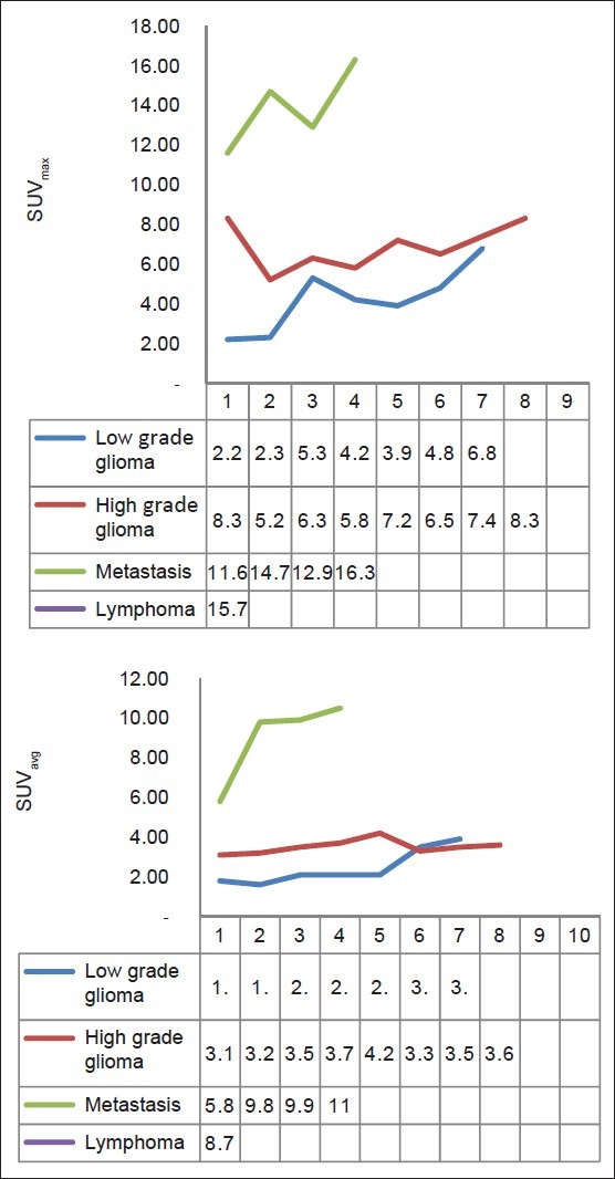
We evaluated 20 patients with an enhancing brain tumor on magnetic resonance imaging (MRI). FDG PET scan was done in all patients pre operatively. For PET image analysis, regions of interest were placed over the tumor (T), contralateral cortex (C), and white matter (WM). Average and maximum pixel values were determined at each site. On the basis of these measurements, average and maximum standard uptake values (SUV avg and SUV max ) were calculated, and comparisons among lesions were then made.

RESULTS

SUVavg and SUVmax are significantly higher for central nervous system (CNS) lymphoma than for other tumors (P < 0.01). High-grade gliomas showed significantly higher SUVavg and SUVmax than the low grade gliomas (P < 0.05) and metastatic tumor showed higher SUVavg and SUVmax than all gliomas, both low and high grade (P < 0.05). When the lowest values of CNS lymphoma parameter were used as cutoff levels to distinguish CNS lymphomas from other tumors (i.e. 100% sensitivity), SUVmax was the most accurate parameter. Using a SUVmax of 15.0 as a cutoff for diagnosing CNS lymphoma, only one case of metastasis (SUV max , 16.3) was found to be false positive in this study.

CONCLUSION

FDG PET appears to provide additional information for differentiating common enhancing malignant brain tumors, namely lymphoma versus high grade glioma and metastatic tumor, particularly when differential diagnoses are difficult to narrow using MRI alone.

摘要

目的

确定F-18-氟脱氧葡萄糖正电子发射断层扫描(F-18-FDG PET)是否可用于区分常见的强化脑肿瘤,如胶质瘤、脑转移瘤和淋巴瘤。

材料与方法

我们评估了20例磁共振成像(MRI)显示有强化脑肿瘤的患者。所有患者术前均进行了FDG PET扫描。对于PET图像分析,在肿瘤(T)、对侧皮质(C)和白质(WM)上放置感兴趣区。在每个部位确定平均像素值和最大像素值。基于这些测量结果,计算平均标准摄取值(SUV avg)和最大标准摄取值(SUV max),然后对病变进行比较。

结果

中枢神经系统(CNS)淋巴瘤的SUVavg和SUVmax显著高于其他肿瘤(P < 0.01)。高级别胶质瘤的SUVavg和SUVmax显著高于低级别胶质瘤(P < 0.05),转移瘤的SUVavg和SUVmax高于所有低级别和高级别胶质瘤(P < 0.05)。当将CNS淋巴瘤参数的最低值用作区分CNS淋巴瘤与其他肿瘤的临界值水平时(即100%敏感性),SUVmax是最准确的参数。在本研究中,以SUVmax为15.0作为诊断CNS淋巴瘤的临界值时,仅发现1例转移瘤(SUV max为16.3)为假阳性。

结论

FDG PET似乎为区分常见的强化恶性脑肿瘤,即淋巴瘤与高级别胶质瘤和转移瘤,提供了额外信息,特别是在仅使用MRI难以缩小鉴别诊断范围时。

https://cdn.ncbi.nlm.nih.gov/pmc/blobs/b0d4/3613621/e980b76fe429/IJNM-26-171-g004.jpg
https://cdn.ncbi.nlm.nih.gov/pmc/blobs/b0d4/3613621/83e6fc525d31/IJNM-26-171-g002.jpg
https://cdn.ncbi.nlm.nih.gov/pmc/blobs/b0d4/3613621/191146a6c669/IJNM-26-171-g003.jpg
https://cdn.ncbi.nlm.nih.gov/pmc/blobs/b0d4/3613621/e980b76fe429/IJNM-26-171-g004.jpg
https://cdn.ncbi.nlm.nih.gov/pmc/blobs/b0d4/3613621/83e6fc525d31/IJNM-26-171-g002.jpg
https://cdn.ncbi.nlm.nih.gov/pmc/blobs/b0d4/3613621/191146a6c669/IJNM-26-171-g003.jpg
https://cdn.ncbi.nlm.nih.gov/pmc/blobs/b0d4/3613621/e980b76fe429/IJNM-26-171-g004.jpg

相似文献

1
Role of (18)F-fluorodeoxyglucose Positron Emission Tomography scan in differentiating enhancing brain tumors.(18)F-氟脱氧葡萄糖正电子发射断层扫描在鉴别强化脑肿瘤中的作用
Indian J Nucl Med. 2011 Oct;26(4):171-6. doi: 10.4103/0972-3919.106698.
2
18F-FDG PET of common enhancing malignant brain tumors.常见强化型恶性脑肿瘤的18F-FDG PET检查
AJR Am J Roentgenol. 2008 Jun;190(6):W365-9. doi: 10.2214/AJR.07.2660.
3
More advantages in detecting bone and soft tissue metastases from prostate cancer using F-PSMA PET/CT.使用F-PSMA PET/CT检测前列腺癌骨和软组织转移方面有更多优势。
Hell J Nucl Med. 2019 Jan-Apr;22(1):6-9. doi: 10.1967/s002449910952. Epub 2019 Mar 7.
4
Common malignant brain tumors: can 18F-FDG PET/CT aid in differentiation?常见恶性脑肿瘤:18F-FDG PET/CT 能否有助于鉴别诊断?
Nucl Med Commun. 2017 Dec;38(12):1109-1116. doi: 10.1097/MNM.0000000000000753.
5
Can the standardized uptake value characterize primary brain tumors on FDG-PET?
Eur J Nucl Med. 1999 Nov;26(11):1501-9. doi: 10.1007/s002590050487.
6
A new diagnostic marker for differentiating multicentric gliomas from multiple intracranial diffuse large B-cell lymphomas on 18F-FDG PET images.一种用于在18F-FDG PET图像上区分多中心胶质瘤与多发颅内弥漫性大B细胞淋巴瘤的新型诊断标志物。
Medicine (Baltimore). 2017 Aug;96(32):e7756. doi: 10.1097/MD.0000000000007756.
7
F-FDG PET/CT in immunocompetent patients with primary central nervous system lymphoma: Differentiation from glioblastoma and correlation with DWI.免疫功能正常的原发性中枢神经系统淋巴瘤患者的 F-FDG PET/CT:与胶质母细胞瘤的鉴别诊断及与 DWI 的相关性。
Eur J Radiol. 2018 Jul;104:26-32. doi: 10.1016/j.ejrad.2018.04.020. Epub 2018 Apr 22.
8
Imaging proliferation in brain tumors with 18F-FLT PET: comparison with 18F-FDG.利用18F-FLT PET成像检测脑肿瘤增殖:与18F-FDG的比较。
J Nucl Med. 2005 Jun;46(6):945-52.
9
¹¹C-methionine and ¹⁸F-fluorodeoxyglucose positron emission tomography/CT in the evaluation of patients with suspected primary and residual/recurrent gliomas.¹¹C-蛋氨酸和 ¹⁸F-氟代脱氧葡萄糖正电子发射断层扫描/计算机断层扫描在疑似原发性和残留/复发性脑胶质瘤患者中的评估。
Chin Med J (Engl). 2012 Jan;125(1):91-6.
10
Comparison of effects of 18F-FDG PET-CT and MRI in identifying and grading gliomas.18F-FDG PET-CT与MRI在胶质瘤识别及分级中的效果比较。
J Biol Regul Homeost Agents. 2016 Jul-Sep;30(3):833-838.

引用本文的文献

1
Predictors for the Differentiation between Glioblastoma, Primary Central Nervous System Lymphoma, and Metastasis in Patients with a Solitary Enhancing Intracranial Mass.孤立性颅内强化肿块患者中胶质母细胞瘤、原发性中枢神经系统淋巴瘤和转移瘤鉴别的预测因素
Asian J Neurosurg. 2024 Jun 6;19(2):186-201. doi: 10.1055/s-0044-1787051. eCollection 2024 Jun.
2
Metabolic brain imaging with glucosamine CEST MRI: in vivo characterization and first insights.使用葡糖胺 CEST MRI 进行代谢脑成像:体内特征描述和初步见解。
Sci Rep. 2023 Dec 12;13(1):22030. doi: 10.1038/s41598-023-48515-5.
3
Correlation Between F-FDG Uptake and Immune Cell Infiltration in Metastatic Brain Lesions.

本文引用的文献

1
18F-FDG PET of common enhancing malignant brain tumors.常见强化型恶性脑肿瘤的18F-FDG PET检查
AJR Am J Roentgenol. 2008 Jun;190(6):W365-9. doi: 10.2214/AJR.07.2660.
2
Evaluation of different cerebral mass lesions by perfusion-weighted MR imaging.通过灌注加权磁共振成像评估不同的脑实质病变
J Magn Reson Imaging. 2006 Oct;24(4):817-24. doi: 10.1002/jmri.20707.
3
Early change in glucose metabolic rate measured using FDG-PET in patients with high-grade glioma predicts response to temozolomide but not temozolomide plus radiotherapy.
转移性脑病变中F-FDG摄取与免疫细胞浸润的相关性
Front Oncol. 2021 Jun 24;11:618705. doi: 10.3389/fonc.2021.618705. eCollection 2021.
4
A preliminary PET radiomics study of brain metastases using a fully automatic segmentation method.一种使用全自动分割方法的脑转移瘤初步 PET 放射组学研究。
BMC Bioinformatics. 2020 Sep 16;21(Suppl 8):325. doi: 10.1186/s12859-020-03647-7.
5
Utility of flouro-deoxy-glucose positron emission tomography/computed tomography in the diagnostic and staging evaluation of patients with primary CNS lymphoma.氟代脱氧葡萄糖正电子发射断层扫描/计算机断层扫描在原发性中枢神经系统淋巴瘤患者诊断及分期评估中的应用
CNS Oncol. 2019 Dec 1;8(4):CNS46. doi: 10.2217/cns-2019-0016. Epub 2019 Nov 29.
6
Utility of FET-PET in detecting high-grade gliomas presenting with equivocal MR imaging features.氟代乙基酪氨酸正电子发射断层扫描(FET-PET)在检测具有模糊磁共振成像(MR)特征的高级别胶质瘤中的应用价值。
World J Nucl Med. 2019 Jul-Sep;18(3):266-272. doi: 10.4103/wjnm.WJNM_89_18.
7
Magnetic Resonance Imaging-Based Screening for Asymptomatic Brain Tumors: A Review.基于磁共振成像的无症状脑肿瘤筛查:综述。
Oncologist. 2019 Mar;24(3):375-384. doi: 10.1634/theoncologist.2018-0177. Epub 2018 Oct 10.
8
Differential diagnosis of posterior fossa brain tumors: Multiple discriminant analysis of Tl-SPECT and FDG-PET.后颅窝脑肿瘤的鉴别诊断:Tl-SPECT与FDG-PET的多重判别分析
Medicine (Baltimore). 2017 Aug;96(33):e7767. doi: 10.1097/MD.0000000000007767.
使用氟代脱氧葡萄糖正电子发射断层扫描(FDG-PET)测量的高级别胶质瘤患者葡萄糖代谢率的早期变化可预测对替莫唑胺的反应,但不能预测对替莫唑胺加放疗的反应。
Int J Radiat Oncol Biol Phys. 2006 Oct 1;66(2):331-8. doi: 10.1016/j.ijrobp.2006.04.043. Epub 2006 Jul 12.
4
Perfusion and diffusion MR imaging in enhancing malignant cerebral tumors.增强型恶性脑肿瘤的灌注与扩散磁共振成像
Eur J Radiol. 2006 Jun;58(3):394-403. doi: 10.1016/j.ejrad.2005.12.032. Epub 2006 Mar 9.
5
FDG-PET in immunocompetent patients with primary central nervous system lymphoma: correlation with MRI and clinical follow-up.免疫功能正常的原发性中枢神经系统淋巴瘤患者的氟代脱氧葡萄糖正电子发射断层扫描(FDG-PET):与磁共振成像(MRI)及临床随访的相关性
Eur J Nucl Med Mol Imaging. 2006 Feb;33(2):164-8. doi: 10.1007/s00259-005-1917-6. Epub 2005 Oct 12.
6
The use of positron-emitting radioisotopes for the localization of brain tumors.
Science. 1951 May 4;113(2940):525-7. doi: 10.1126/science.113.2940.525.
7
Imaging gliomas with positron emission tomography and single-photon emission computed tomography.利用正电子发射断层扫描和单光子发射计算机断层扫描对胶质瘤进行成像。
Semin Nucl Med. 2003 Apr;33(2):148-62. doi: 10.1053/snuc.2003.127304.
8
PET in lymphoma: what are the oncologist's needs?淋巴瘤中的正电子发射断层扫描(PET):肿瘤学家的需求是什么?
Eur J Nucl Med Mol Imaging. 2003 Jun;30 Suppl 1:S37-41. doi: 10.1007/s00259-003-1158-5. Epub 2003 Apr 4.
9
Combination of single-voxel proton MR spectroscopy and apparent diffusion coefficient calculation in the evaluation of common brain tumors.单体素质子磁共振波谱与表观扩散系数计算相结合在常见脑肿瘤评估中的应用
AJNR Am J Neuroradiol. 2003 Feb;24(2):225-33.
10
Can the standardized uptake value characterize primary brain tumors on FDG-PET?
Eur J Nucl Med. 1999 Nov;26(11):1501-9. doi: 10.1007/s002590050487.