• 文献检索
  • 文档翻译
  • 深度研究
  • 学术资讯
  • Suppr Zotero 插件Zotero 插件
  • 邀请有礼
  • 套餐&价格
  • 历史记录
应用&插件
Suppr Zotero 插件Zotero 插件浏览器插件Mac 客户端Windows 客户端微信小程序
定价
高级版会员购买积分包购买API积分包
服务
文献检索文档翻译深度研究API 文档MCP 服务
关于我们
关于 Suppr公司介绍联系我们用户协议隐私条款
关注我们

Suppr 超能文献

核心技术专利:CN118964589B侵权必究
粤ICP备2023148730 号-1Suppr @ 2026

文献检索

告别复杂PubMed语法,用中文像聊天一样搜索,搜遍4000万医学文献。AI智能推荐,让科研检索更轻松。

立即免费搜索

文件翻译

保留排版,准确专业,支持PDF/Word/PPT等文件格式,支持 12+语言互译。

免费翻译文档

深度研究

AI帮你快速写综述,25分钟生成高质量综述,智能提取关键信息,辅助科研写作。

立即免费体验

转化生长因子β在结节硬化型霍奇金淋巴瘤发病机制中作用的免疫组化证据

Immunohistochemical evidence of a role for transforming growth factor beta in the pathogenesis of nodular sclerosing Hodgkin's disease.

作者信息

Kadin M E, Agnarsson B A, Ellingsworth L R, Newcom S R

机构信息

Department of Pathology, Beth Israel Hospital, Boston, Massachusetts 02215.

出版信息

Am J Pathol. 1990 Jun;136(6):1209-14.

PMID:2356855
原文链接:https://pmc.ncbi.nlm.nih.gov/articles/PMC1877568/
Abstract

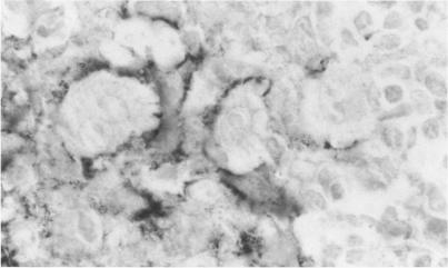
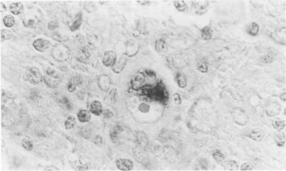
Transforming growth factor beta (TFG-beta) is a multifunctional growth factor that promotes the growth of fibroblasts, collagen synthesis and angiogenesis, and stimulates monocyte migration and activation, but suppresses the growth and differentiation of immune lymphocytes and killer cells. Previously we demonstrated biologic activity for TGF-beta in supernatants of fresh Hodgkin's disease (HD) cell cultures and the cell line L428 derived from nodular sclerosing HD. This study was undertaken to find evidence of TGF-beta activity directly in tissues affected by HD. Formalin-fixed tissue from 14 patients with HD, including 8 nodular sclerosis, 4 mixed cellularity, 1 lymphocyte predominance, and 1 lymphocyte depletion type were studied by immunoperoxidase technique with antibody CC (1-30) raised against a synthetic polypeptide with the same N-terminal amino acid sequence as TGF-beta 1. Transforming growth factor beta activity was demonstrated in six cases of nodular sclerosis but not in other histologic types of HD. Staining for TGF-beta was found in the cytoplasm of Reed-Sternberg (RS) cells in one case and on the surface of RS cells and their lacunar variants in five cases. Transforming growth factor beta activity associated with the extracellular matrix was localized mainly around blood vessels, zones of necrosis, at the margins of bands of collagen sclerosis, and in areas containing syncytia of RS cells. In two cases TGF-beta was associated with collections of epithelioid histiocytes or granulomas. Small lymphocytes, granulocytes, and germinal center cells were unreactive. These results suggest that TGF-beta is a growth factor of biologic importance in HD and may be responsible for many of the histologic features, such as nodular sclerosis and granulomas, that may have prognostic significance.

摘要

转化生长因子β(TGF-β)是一种多功能生长因子,可促进成纤维细胞生长、胶原蛋白合成和血管生成,刺激单核细胞迁移和活化,但会抑制免疫淋巴细胞和杀伤细胞的生长与分化。此前我们已证明新鲜霍奇金淋巴瘤(HD)细胞培养上清液及源自结节硬化型HD的L428细胞系中存在TGF-β的生物活性。本研究旨在直接在受HD影响的组织中寻找TGF-β活性的证据。采用免疫过氧化物酶技术,使用针对与TGF-β1具有相同N端氨基酸序列的合成多肽产生的抗体CC(1-30),对14例HD患者的福尔马林固定组织进行研究,其中包括8例结节硬化型、4例混合细胞型、1例淋巴细胞为主型和1例淋巴细胞消减型。在6例结节硬化型病例中检测到转化生长因子β活性,而在其他组织学类型的HD中未检测到。在1例病例的里德-斯腾伯格(RS)细胞胞质中发现TGF-β染色,在5例病例的RS细胞及其陷窝变体表面发现TGF-β染色。与细胞外基质相关的转化生长因子β活性主要定位于血管周围、坏死区域、胶原硬化带边缘以及含有RS细胞合体的区域。在2例病例中,TGF-β与上皮样组织细胞或肉芽肿聚集有关。小淋巴细胞、粒细胞和生发中心细胞无反应。这些结果表明,TGF-β是HD中具有生物学重要性的生长因子,可能是许多具有预后意义的组织学特征(如结节硬化和肉芽肿)的原因。

https://cdn.ncbi.nlm.nih.gov/pmc/blobs/1c74/1877568/46ade7e48d27/amjpathol00114-0019-b.jpg
https://cdn.ncbi.nlm.nih.gov/pmc/blobs/1c74/1877568/400017d5e350/amjpathol00114-0018-a.jpg
https://cdn.ncbi.nlm.nih.gov/pmc/blobs/1c74/1877568/2dbf0488c6c4/amjpathol00114-0018-b.jpg
https://cdn.ncbi.nlm.nih.gov/pmc/blobs/1c74/1877568/a42600499654/amjpathol00114-0019-a.jpg
https://cdn.ncbi.nlm.nih.gov/pmc/blobs/1c74/1877568/46ade7e48d27/amjpathol00114-0019-b.jpg

相似文献

1
Immunohistochemical evidence of a role for transforming growth factor beta in the pathogenesis of nodular sclerosing Hodgkin's disease.转化生长因子β在结节硬化型霍奇金淋巴瘤发病机制中作用的免疫组化证据
Am J Pathol. 1990 Jun;136(6):1209-14.
2
Expression of T-cell antigens on Reed-Sternberg cells in a subset of patients with nodular sclerosing and mixed cellularity Hodgkin's disease.在部分结节硬化型和混合细胞型霍奇金淋巴瘤患者中,里德-斯腾伯格细胞上T细胞抗原的表达情况。
Am J Pathol. 1988 Feb;130(2):345-53.
3
Eosinophils are the major source of transforming growth factor-beta 1 in nodular sclerosing Hodgkin's disease.嗜酸性粒细胞是结节硬化型霍奇金淋巴瘤中转化生长因子-β1的主要来源。
Am J Pathol. 1993 Jan;142(1):11-6.
4
High molecular weight transforming growth factor beta is excreted in the urine in active nodular sclerosing Hodgkin's disease.
Cancer Res. 1992 Dec 15;52(24):6768-73.
5
Hodgkin's disease, lymphocyte predominance type, nodular--further evidence for a B cell derivation. L & H variants of Reed-Sternberg cells express L26, a pan B cell marker.霍奇金淋巴瘤,淋巴细胞为主型,结节性——B细胞起源的进一步证据。里德-施特恩贝格细胞的L&H变异型表达L26,一种泛B细胞标志物。
Am J Pathol. 1988 Nov;133(2):211-7.
6
Fascin, a sensitive new marker for Reed-Sternberg cells of hodgkin's disease. Evidence for a dendritic or B cell derivation?Fascin,一种霍奇金病里德-斯腾伯格细胞的新型敏感标志物。它来源于树突状细胞还是B细胞?
Am J Pathol. 1997 Feb;150(2):543-62.
7
Hodgkin's disease, lymphocyte predominance type, nodular--a distinct entity? Unique staining profile for L&H variants of Reed-Sternberg cells defined by monoclonal antibodies to leukocyte common antigen, granulocyte-specific antigen, and B-cell-specific antigen.霍奇金病,淋巴细胞为主型,结节性——一种独特的实体?通过针对白细胞共同抗原、粒细胞特异性抗原和B细胞特异性抗原的单克隆抗体确定的里德-斯腾伯格细胞L&H变异型的独特染色特征。
Am J Pathol. 1985 Jan;118(1):1-6.
8
Immunohistochemical detection of Epstein-Barr virus-encoded latent membrane protein in Reed-Sternberg cells and variants of Hodgkin's disease.霍奇金病的里德-斯腾伯格细胞及其变异型中爱泼斯坦-巴尔病毒编码的潜伏膜蛋白的免疫组织化学检测
Mod Pathol. 1994 May;7(4):454-61.
9
Macrophage-derived chemokine expression in classical Hodgkin's lymphoma: application of tissue microarrays.
Mod Pathol. 2001 Dec;14(12):1270-6. doi: 10.1038/modpathol.3880473.
10
Vascular endothelial growth factor (VEGF) is expressed by neoplastic Hodgkin-Reed-Sternberg cells in Hodgkin's disease.血管内皮生长因子(VEGF)在霍奇金病的肿瘤性霍奇金-里德-斯腾伯格细胞中表达。
J Pathol. 2002 Aug;197(5):677-83. doi: 10.1002/path.1151.

引用本文的文献

1
Successful Treatment of Childhood Hodgkin Lymphoma and Secondary Myelofibrosis Resistant to Intensive Therapy, Including Allogeneic Transplantation.成功治疗对强化治疗(包括异基因移植)耐药的儿童霍奇金淋巴瘤和继发性骨髓纤维化
Turk J Haematol. 2025 May 22;42(2):158-160. doi: 10.4274/tjh.galenos.2025.2025.0044. Epub 2025 Apr 7.
2
The Tumor Microenvironment in Classic Hodgkin's Lymphoma in Responder and No-Responder Patients to First Line ABVD Therapy.一线ABVD治疗有反应和无反应的经典霍奇金淋巴瘤患者的肿瘤微环境
Cancers (Basel). 2023 May 17;15(10):2803. doi: 10.3390/cancers15102803.
3
Filling the Gap: The Immune Therapeutic Armamentarium for Relapsed/Refractory Hodgkin Lymphoma.

本文引用的文献

1
Impact of Hodgkin's disease upon the immune system.霍奇金淋巴瘤对免疫系统的影响。
Semin Oncol. 1980 Jun;7(2):114-25.
2
Potentiation of fibroblast growth by nodular sclerosing Hodgkin's disease cell cultures.结节性硬化型霍奇金病细胞培养对成纤维细胞生长的增强作用。
Blood. 1982 Jul;60(1):228-37.
3
Clinicopathologic studies of 117 untreated patients subjected to laparotomy for the staging of Hodgkin's disease.对117例未经治疗的霍奇金淋巴瘤患者进行剖腹手术分期的临床病理研究。
填补空白:复发/难治性霍奇金淋巴瘤的免疫治疗武器库
J Clin Med. 2022 Nov 6;11(21):6574. doi: 10.3390/jcm11216574.
4
Inflammatory microenvironment in classical Hodgkin's lymphoma with special stress on mast cells.经典型霍奇金淋巴瘤中的炎性微环境,特别强调肥大细胞。
Front Oncol. 2022 Oct 13;12:964573. doi: 10.3389/fonc.2022.964573. eCollection 2022.
5
Histological Subtypes Drive Distinct Prognostic Immune Signatures in Classical Hodgkin Lymphoma.组织学亚型决定经典型霍奇金淋巴瘤独特的预后免疫特征。
Cancers (Basel). 2022 Oct 6;14(19):4893. doi: 10.3390/cancers14194893.
6
Formation of the Immunosuppressive Microenvironment of Classic Hodgkin Lymphoma and Therapeutic Approaches to Counter It.经典型霍奇金淋巴瘤免疫抑制微环境的形成及应对治疗策略。
Int J Mol Sci. 2019 May 15;20(10):2416. doi: 10.3390/ijms20102416.
7
The Dynamic Roles of TGF-β Signalling in EBV-Associated Cancers.转化生长因子-β信号在EB病毒相关癌症中的动态作用
Cancers (Basel). 2018 Jul 27;10(8):247. doi: 10.3390/cancers10080247.
8
Insights into the molecular roles of heparan sulfate proteoglycans (HSPGs-syndecans) in autocrine and paracrine growth factor signaling in the pathogenesis of Hodgkin's lymphoma.硫酸乙酰肝素蛋白聚糖(HSPGs-多配体蛋白聚糖)在霍奇金淋巴瘤发病机制中的自分泌和旁分泌生长因子信号传导中的分子作用研究
Tumour Biol. 2016 Sep;37(9):11573-11588. doi: 10.1007/s13277-016-5118-7. Epub 2016 Jun 18.
9
Role of mast cells in fibrosis of classical Hodgkin lymphoma.肥大细胞在经典型霍奇金淋巴瘤纤维化中的作用
Int J Immunopathol Pharmacol. 2016 Dec;29(4):603-611. doi: 10.1177/0394632016644447. Epub 2016 Apr 19.
10
Hodgkin's lymphoma associated with myelofibrosis: A case report.霍奇金淋巴瘤合并骨髓纤维化:一例报告。
Oncol Lett. 2015 Sep;10(3):1551-1554. doi: 10.3892/ol.2015.3438. Epub 2015 Jun 30.
Cancer. 1971 Jun;27(6):1277-94. doi: 10.1002/1097-0142(197106)27:6<1277::aid-cncr2820270602>3.0.co;2-t.
4
The Hodgkin's cell in nodular sclerosis does not release interleukin-1.结节性硬化型中的霍奇金细胞不释放白细胞介素-1。
J Lab Clin Med. 1985 Feb;105(2):170-7.
5
Hepatic processing of transforming growth factor beta in the rat. Uptake, metabolism, and biliary excretion.大鼠中转化生长因子β的肝脏处理。摄取、代谢及胆汁排泄。
J Clin Invest. 1987 Sep;80(3):750-7. doi: 10.1172/JCI113130.
6
Synthesis of messenger RNAs for transforming growth factors alpha and beta and the epidermal growth factor receptor by human tumors.人类肿瘤中转化生长因子α和β以及表皮生长因子受体信使核糖核酸的合成
Cancer Res. 1987 Feb 1;47(3):707-12.
7
Hodgkin's disease, lymphocyte predominance type, nodular--further evidence for a B cell derivation. L & H variants of Reed-Sternberg cells express L26, a pan B cell marker.霍奇金淋巴瘤,淋巴细胞为主型,结节性——B细胞起源的进一步证据。里德-施特恩贝格细胞的L&H变异型表达L26,一种泛B细胞标志物。
Am J Pathol. 1988 Nov;133(2):211-7.
8
The glioblastoma-derived T-cell suppressor factor/transforming growth factor beta 2 inhibits the generation of lymphokine-activated killer (LAK) cells.胶质母细胞瘤衍生的T细胞抑制因子/转化生长因子β2可抑制淋巴因子激活的杀伤细胞(LAK细胞)的生成。
Int J Cancer. 1988 Oct 15;42(4):562-7. doi: 10.1002/ijc.2910420416.
9
L-428 Reed-Sternberg cells and mononuclear Hodgkin's cells arise from a single cloned mononuclear cell.L-428里德-斯腾伯格细胞和单核霍奇金细胞源自单个克隆的单核细胞。
Int J Cell Cloning. 1988 Nov;6(6):417-31. doi: 10.1002/stem.5530060606.
10
Effects of transforming growth factor-beta on human lymphokine-activated killer cell precursors. Autocrine inhibition of cellular proliferation and differentiation to immune killer cells.转化生长因子-β对人淋巴因子激活的杀伤细胞前体的作用。对细胞增殖和向免疫杀伤细胞分化的自分泌抑制。
J Immunol. 1988 Jul 15;141(2):690-8.