Pinkus G S, Pinkus J L, Langhoff E, Matsumura F, Yamashiro S, Mosialos G, Said J W
Department of Pathology, Brigham and Women's Hospital, Boston, MA 02115, USA.
Am J Pathol. 1997 Feb;150(2):543-62.
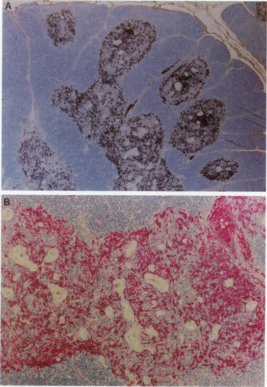
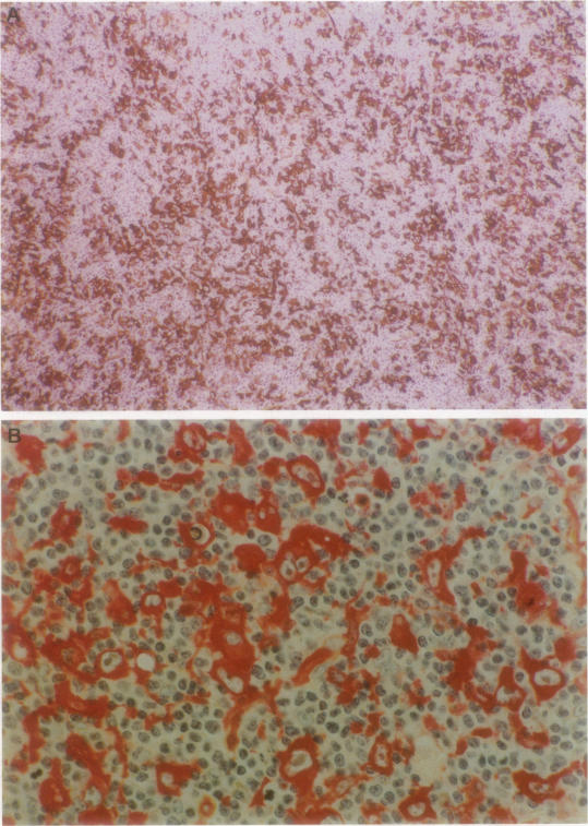
Immunohistochemical localization of human fascin, a distinct 55-kd actin-bundling protein, was determined for a wide variety of lymphoid tissues (364 specimens total). In non-neoplastic tissues, reactivity was highly selective and localized predominantly in dendritic cells. In the thymus, this protein was distinctly localized to medullary dendritic cells. In reactive nodes, interdigitating reticulum cells of T zones, cells in subcapsular areas, and cells of the reticular network were reactive, with variable reactivity observed for follicular dendritic cells. Splenic dendritic cells of the white pulp and sinus-lining cells of the red pulp were reactive. Endothelial cells of all tissues exhibited variable reactivity. Lymphoid cells, myeloid cells, and plasma cells were uniformly nonreactive. In the peripheral blood, only dendritic (veiled) cells were reactive for fascin. A striking finding was observed for cases of Hodgkin's disease (total 187 cases). In all cases of nodular sclerosis (132), mixed cellularity (34), lymphocyte depletion (2), and unclassified types (5), all or nearly all Reed-Sternberg cells and variants were immunoreactive for fascin. Neoplastic cells exhibited strong diffuse cytoplasmic staining and frequently assumed dendritic shapes, particularly in the nodular sclerosis type, producing an interdigitating meshwork or syncytial network of cells. In cases of mixed cellularity type, neoplastic cells generally appeared more discrete. In all 14 cases of nodular lymphocyte predominance type, L&H variants were nonreactive. By contrast, neoplastic lymphoid cells of only 24 of 156 (15%) other lymphoid neoplasms (127 B cell, 27 T cell, and two null cell evaluated) were reactive for fascin. Fascin represents a highly effective marker for detection of certain dendritic cells in normal and neoplastic tissues, is an extremely consistent marker for Reed-Sternberg cells and variants of Hodgkin's disease (except L&H types), and may be helpful to distinguish between Hodgkin's disease and non-Hodgkin's lymphoma in difficult cases. The staining profile for fascin raises the possibility of a dendritic cell derivation, particularly an interdigitating reticulum cell, for the neoplastic cells of Hodgkin's disease, notably in nodular sclerosis type. However, as fascin expression may be induced by Epstein-Barr virus infection of B cells, the possibility that viral induction of fascin in lymphoid or other cell types must also be considered in Epstein-Barr virus-positive cases.
对大量淋巴组织(共364个标本)进行了人丝聚蛋白(一种独特的55-kd肌动蛋白成束蛋白)的免疫组织化学定位。在非肿瘤组织中,反应具有高度选择性,主要定位于树突状细胞。在胸腺中,该蛋白明显定位于髓质树突状细胞。在反应性淋巴结中,T区的交错网状细胞、被膜下区域的细胞以及网状网络的细胞呈反应性,滤泡树突状细胞的反应性各不相同。白髓的脾树突状细胞和红髓的窦衬细胞呈反应性。所有组织的内皮细胞均表现出不同程度的反应性。淋巴细胞、髓细胞和浆细胞均无反应性。在外周血中,只有树突状(面纱样)细胞对丝聚蛋白呈反应性。在霍奇金病病例(共187例)中观察到一个显著发现。在所有结节硬化型(132例)、混合细胞型(34例)、淋巴细胞消减型(2例)和未分类型(5例)中,所有或几乎所有的里德-斯腾伯格细胞及其变异型对丝聚蛋白均呈免疫反应性。肿瘤细胞表现出强烈的弥漫性胞质染色,且常呈树突状形态,尤其是在结节硬化型中,形成细胞交错网络或多核细胞网络。在混合细胞型病例中,肿瘤细胞通常显得更为离散。在所有14例结节性淋巴细胞为主型病例中,L&H变异型无反应性。相比之下,在156例其他淋巴肿瘤(评估了127例B细胞、27例T细胞和2例裸细胞)中,只有24例(15%)的肿瘤性淋巴细胞对丝聚蛋白呈反应性。丝聚蛋白是检测正常和肿瘤组织中某些树突状细胞的高效标志物,是霍奇金病里德-斯腾伯格细胞及其变异型(L&H型除外)极为一致的标志物,在疑难病例中可能有助于区分霍奇金病和非霍奇金淋巴瘤。丝聚蛋白染色模式提示霍奇金病的肿瘤细胞,尤其是结节硬化型,可能起源于树突状细胞,特别是交错网状细胞。然而,由于丝聚蛋白表达可能由B细胞的爱泼斯坦-巴尔病毒感染诱导,在爱泼斯坦-巴尔病毒阳性病例中也必须考虑淋巴或其他细胞类型中病毒诱导丝聚蛋白表达的可能性。