Department of Internal Medicine, Erasmus MC, University Medical Center Rotterdam, Rotterdam, The Netherlands.
PLoS One. 2013;8(4):e60784. doi: 10.1371/journal.pone.0060784. Epub 2013 Apr 1.
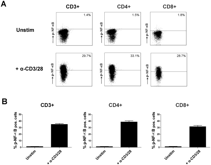
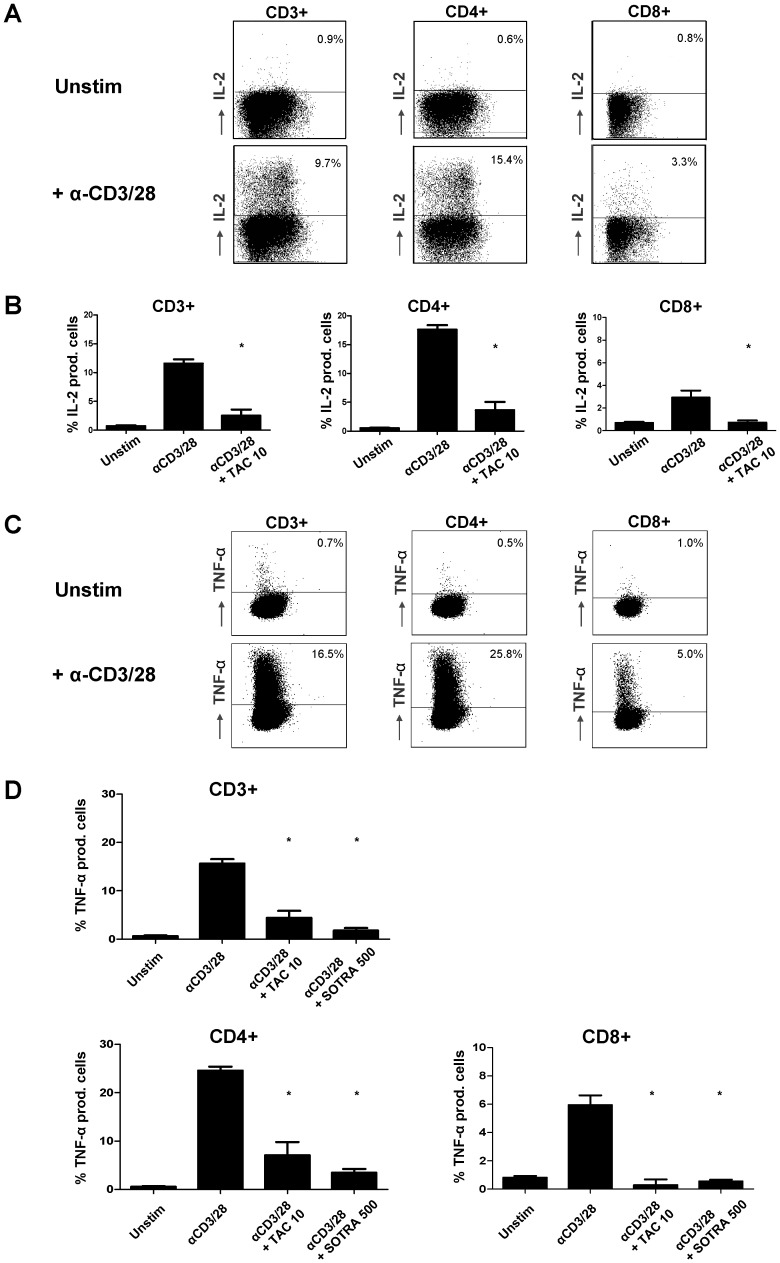
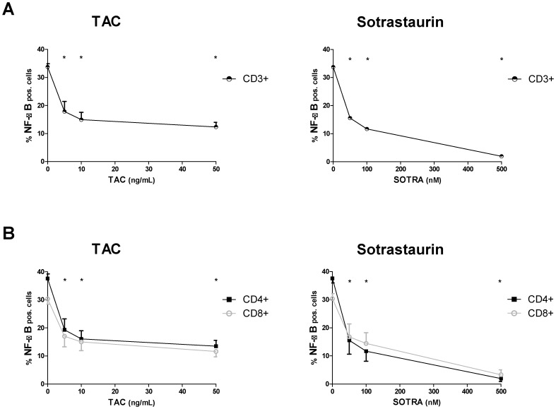
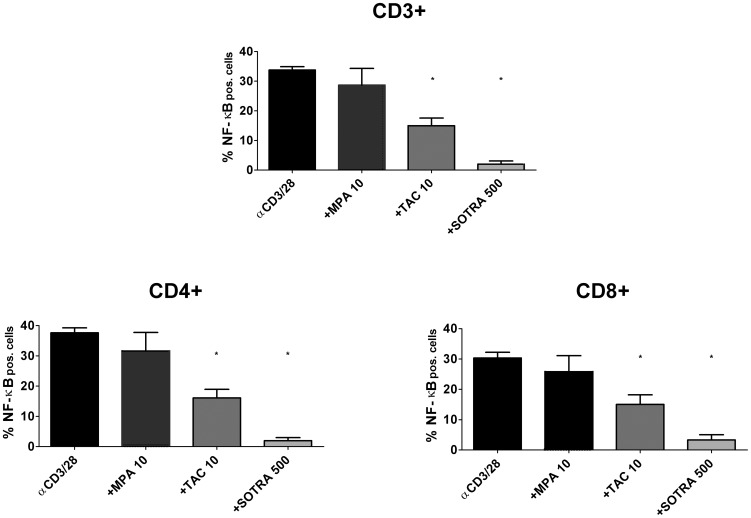
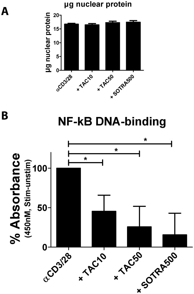
The calcineurin inhibitor, tacrolimus (TAC), inhibits the protein phosphatase activity of calcineurin, leading to suppression of the nuclear translocation of NFAT and inhibition of T cell activation. Apart from NFAT also the transcription factor NF-κB plays a key functional role in T cell activation. Therefore, blockade of the NF-κB activation cascade by immunosuppressive drugs prevents immune activation. Here we studied whether TAC blocks NF-κB activation in peripheral human T cells. After anti-CD3/CD28-activation of T cells from healthy volunteers, NF-κB (p65) phosphorylation was measured by flow cytometry in CD3+ T cells, CD4+ helper T cells and CD8+ cytotoxic T cells in the absence and presence of TAC 10 ng/mL, sotrastaurin 500 nM (positive control) and mycophenolic acid 10 µg/mL (negative control; n = 6). NF-κB transcriptional activity was measured by ELISA and intracellular TNFα protein, a downstream target, was measured by flow cytometry to assess the functional consequences of NF-κB blockade. Anti-CD3/28-activation induced NF-κB phosphorylation in CD3+ T cells, CD4+ T cells and CD8+ T cells by 34% (mean), 38% and 30% resp. (p<0.01). Sotrastaurin inhibited NF-κB activation in the respective T cell subsets by 93%, 95% and 86% (p<0.01 vs. no drug), while mycophenolic acid did not affect this activation pathway. Surprisingly, TAC also inhibited NF-κB phosphorylation, by 55% (p<0.01) in CD3+ T cells, by 56% (p<0.01) in CD4+ T cells and by 51% in CD8+ T cells (p<0.01). In addition, TAC suppressed NF-κB DNA binding capacity by 55% (p<0.05) in CD3+ T cells and TNFα protein expression was inhibited in CD3+ T cells, CD4+ T cells and CD8+ T cells by 76%, 71% and 93% resp. (p<0.01 vs. no drug), confirming impaired NF-κB signaling. This study shows the suppressive effect of TAC on NF-κB signaling in peripheral human T cell subsets, measured at three specific positions in the NF-κB activation cascade.
钙调磷酸酶抑制剂他克莫司(TAC)抑制钙调磷酸酶的蛋白磷酸酶活性,导致 NFAT 核易位和 T 细胞活化受到抑制。除了 NFAT 之外,转录因子 NF-κB 在 T 细胞活化中也起着关键的功能作用。因此,免疫抑制剂阻断 NF-κB 激活级联反应可防止免疫激活。在这里,我们研究了 TAC 是否会阻断外周血人 T 细胞中的 NF-κB 激活。在来自健康志愿者的 T 细胞经抗 CD3/CD28 激活后,通过流式细胞术在 CD3+T 细胞、CD4+辅助 T 细胞和 CD8+细胞毒性 T 细胞中测量 NF-κB(p65)磷酸化,在存在和不存在 TAC(10ng/mL)、索他拉唑(500nM,阳性对照)和霉酚酸(10μg/mL,阴性对照;n=6)的情况下。通过 ELISA 测量 NF-κB 转录活性,并通过流式细胞术测量细胞内 TNFα 蛋白(下游靶标),以评估 NF-κB 阻断的功能后果。抗 CD3/28 激活诱导 CD3+T 细胞、CD4+T 细胞和 CD8+T 细胞中的 NF-κB 磷酸化分别增加 34%(平均值)、38%和 30%(p<0.01)。索他拉唑抑制各自 T 细胞亚群中的 NF-κB 激活,分别为 93%、95%和 86%(p<0.01 与无药物相比),而霉酚酸不影响此激活途径。令人惊讶的是,TAC 还抑制 NF-κB 磷酸化,CD3+T 细胞中为 55%(p<0.01),CD4+T 细胞中为 56%(p<0.01),CD8+T 细胞中为 51%(p<0.01)。此外,TAC 抑制 CD3+T 细胞中 NF-κB DNA 结合能力 55%(p<0.05),并抑制 CD3+T 细胞、CD4+T 细胞和 CD8+T 细胞中的 TNFα 蛋白表达,分别为 76%、71%和 93%(p<0.01 与无药物相比),证实 NF-κB 信号转导受损。这项研究显示了 TAC 在人外周血 T 细胞亚群中对 NF-κB 信号的抑制作用,这是在 NF-κB 激活级联反应的三个特定位置测量的。