• 文献检索
  • 文档翻译
  • 深度研究
  • 学术资讯
  • Suppr Zotero 插件Zotero 插件
  • 邀请有礼
  • 套餐&价格
  • 历史记录
应用&插件
Suppr Zotero 插件Zotero 插件浏览器插件Mac 客户端Windows 客户端微信小程序
定价
高级版会员购买积分包购买API积分包
服务
文献检索文档翻译深度研究API 文档MCP 服务
关于我们
关于 Suppr公司介绍联系我们用户协议隐私条款
关注我们

Suppr 超能文献

核心技术专利:CN118964589B侵权必究
粤ICP备2023148730 号-1Suppr @ 2026

文献检索

告别复杂PubMed语法,用中文像聊天一样搜索,搜遍4000万医学文献。AI智能推荐,让科研检索更轻松。

立即免费搜索

文件翻译

保留排版,准确专业,支持PDF/Word/PPT等文件格式,支持 12+语言互译。

免费翻译文档

深度研究

AI帮你快速写综述,25分钟生成高质量综述,智能提取关键信息,辅助科研写作。

立即免费体验

调控细胞增殖和染色质解聚过程中深度分辨纳米级结构变化的研究。

Investigation of depth-resolved nanoscale structural changes in regulated cell proliferation and chromatin decondensation.

作者信息

Uttam Shikhar, Bista Rajan K, Staton Kevin, Alexandrov Sergey, Choi Serah, Bakkenist Christopher J, Hartman Douglas J, Brand Randall E, Liu Yang

机构信息

Biomedical and Optical Imaging Laboratory (BOIL), Departments of Medicine and Bioengineering, University of Pittsburgh, Pittsburgh, PA 15213, USA.

出版信息

Biomed Opt Express. 2013 Apr 1;4(4):596-613. doi: 10.1364/BOE.4.000596. Epub 2013 Mar 22.

DOI:10.1364/BOE.4.000596
PMID:23577294
原文链接:https://pmc.ncbi.nlm.nih.gov/articles/PMC3617721/
Abstract

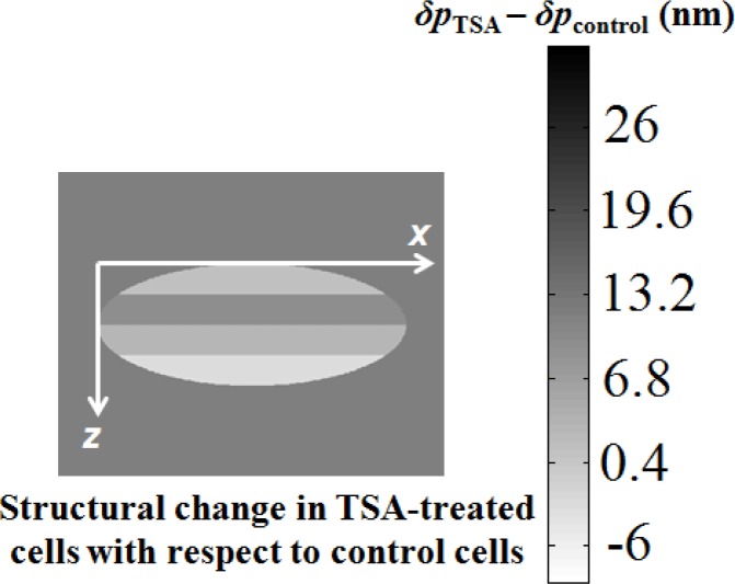
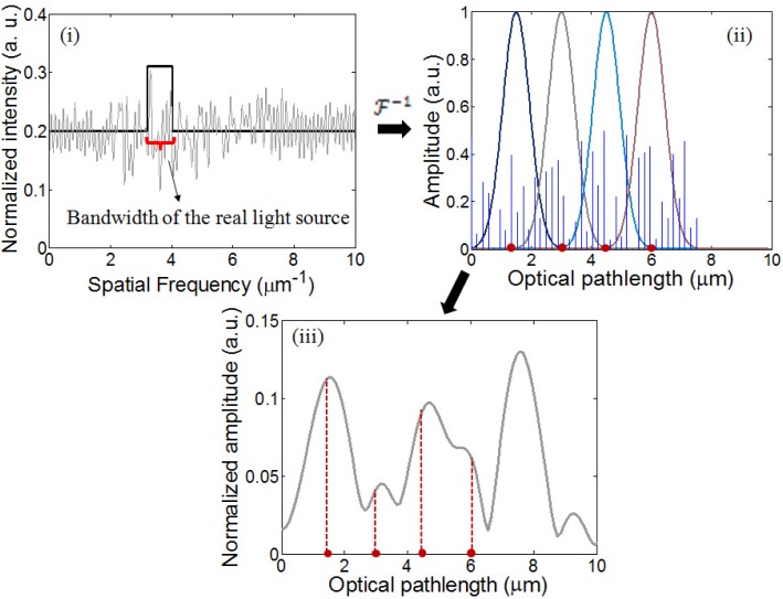
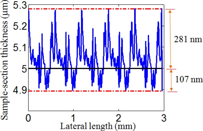
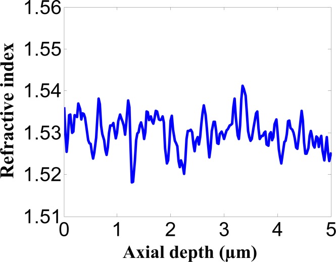
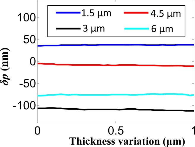
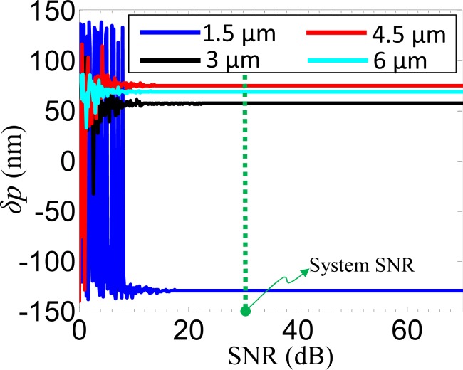
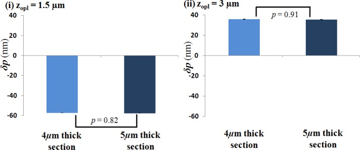
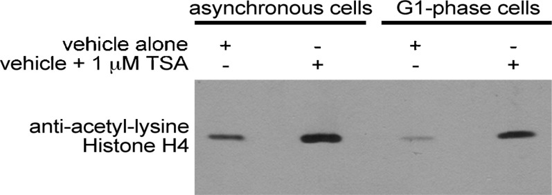
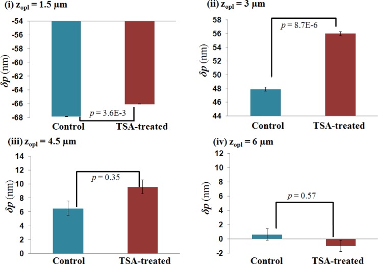
We present depth-resolved spatial-domain low-coherence quantitative phase microscopy, a simple approach that utilizes coherence gating to construct a depth-resolved structural feature vector quantifying sub-resolution axial structural changes at different optical depths within the sample. We show that this feature vector is independent of sample thickness variation, and identifies nanoscale structural changes in clinically prepared samples. We present numerical simulations and experimental validation to demonstrate the feasibility of the approach. We also perform experiments using unstained cells to investigate the nanoscale structural changes in regulated cell proliferation through cell cycle and chromatin decondensation induced by histone acetylation.

摘要

我们提出了深度分辨空间域低相干定量相显微镜技术,这是一种简单的方法,利用相干选通来构建深度分辨结构特征向量,以量化样品内不同光学深度处的亚分辨率轴向结构变化。我们表明,该特征向量与样品厚度变化无关,并能识别临床制备样品中的纳米级结构变化。我们通过数值模拟和实验验证来证明该方法的可行性。我们还使用未染色的细胞进行实验,以研究通过组蛋白乙酰化诱导的细胞周期和染色质解聚在细胞增殖调控中的纳米级结构变化。

https://cdn.ncbi.nlm.nih.gov/pmc/blobs/6b2d/3617721/95448c3f5ff9/boe-4-4-596-g019.jpg
https://cdn.ncbi.nlm.nih.gov/pmc/blobs/6b2d/3617721/72cd2c82a894/boe-4-4-596-g001.jpg
https://cdn.ncbi.nlm.nih.gov/pmc/blobs/6b2d/3617721/19d36ea28412/boe-4-4-596-g002.jpg
https://cdn.ncbi.nlm.nih.gov/pmc/blobs/6b2d/3617721/7e8030bafbda/boe-4-4-596-g003.jpg
https://cdn.ncbi.nlm.nih.gov/pmc/blobs/6b2d/3617721/c91b9008b5de/boe-4-4-596-g004.jpg
https://cdn.ncbi.nlm.nih.gov/pmc/blobs/6b2d/3617721/8e6fd7e9ac31/boe-4-4-596-g005.jpg
https://cdn.ncbi.nlm.nih.gov/pmc/blobs/6b2d/3617721/52c187cfe1d9/boe-4-4-596-g006.jpg
https://cdn.ncbi.nlm.nih.gov/pmc/blobs/6b2d/3617721/0be27e888f10/boe-4-4-596-g007.jpg
https://cdn.ncbi.nlm.nih.gov/pmc/blobs/6b2d/3617721/34499e58e414/boe-4-4-596-g008.jpg
https://cdn.ncbi.nlm.nih.gov/pmc/blobs/6b2d/3617721/ecbe58c7f279/boe-4-4-596-g009.jpg
https://cdn.ncbi.nlm.nih.gov/pmc/blobs/6b2d/3617721/5e9e4d97ccc8/boe-4-4-596-g010.jpg
https://cdn.ncbi.nlm.nih.gov/pmc/blobs/6b2d/3617721/b6898b00447a/boe-4-4-596-g011.jpg
https://cdn.ncbi.nlm.nih.gov/pmc/blobs/6b2d/3617721/f57976906689/boe-4-4-596-g012.jpg
https://cdn.ncbi.nlm.nih.gov/pmc/blobs/6b2d/3617721/3bcf083a7aa0/boe-4-4-596-g013.jpg
https://cdn.ncbi.nlm.nih.gov/pmc/blobs/6b2d/3617721/dfce17cc16ad/boe-4-4-596-g014.jpg
https://cdn.ncbi.nlm.nih.gov/pmc/blobs/6b2d/3617721/e9dbe78fceb8/boe-4-4-596-g015.jpg
https://cdn.ncbi.nlm.nih.gov/pmc/blobs/6b2d/3617721/2b61b661f23f/boe-4-4-596-g016.jpg
https://cdn.ncbi.nlm.nih.gov/pmc/blobs/6b2d/3617721/18b3bf98bada/boe-4-4-596-g017.jpg
https://cdn.ncbi.nlm.nih.gov/pmc/blobs/6b2d/3617721/d6ca6e5cc99c/boe-4-4-596-g018.jpg
https://cdn.ncbi.nlm.nih.gov/pmc/blobs/6b2d/3617721/95448c3f5ff9/boe-4-4-596-g019.jpg

相似文献

1
Investigation of depth-resolved nanoscale structural changes in regulated cell proliferation and chromatin decondensation.调控细胞增殖和染色质解聚过程中深度分辨纳米级结构变化的研究。
Biomed Opt Express. 2013 Apr 1;4(4):596-613. doi: 10.1364/BOE.4.000596. Epub 2013 Mar 22.
2
Erratum: Investigation of depth-resolved nanoscale structural changes in regulated cell proliferation and chromatin decondensation: erratum.勘误:调控细胞增殖和染色质解聚中深度分辨纳米级结构变化的研究:勘误。
Biomed Opt Express. 2013 Oct 14;4(11):2491. doi: 10.1364/BOE.4.002491. eCollection 2013.
3
Fourier phase based depth-resolved nanoscale nuclear architecture mapping for cancer detection.基于傅里叶相位的深度分辨纳米级核结构测绘用于癌症检测。
Methods. 2018 Mar 1;136:134-151. doi: 10.1016/j.ymeth.2017.10.011. Epub 2017 Nov 7.
4
Nano-sensitive optical coherence tomography.纳米敏感染光相干断层扫描技术。
Nanoscale. 2014 Apr 7;6(7):3545-9. doi: 10.1039/c3nr06132a.
5
Accessing depth-resolved high spatial frequency content from the optical coherence tomography signal.从光相干断层扫描信号中获取深度分辨的高空间频率内容。
Sci Rep. 2021 Aug 24;11(1):17123. doi: 10.1038/s41598-021-96619-7.
6
Fourier phase in Fourier-domain optical coherence tomography.傅里叶域光学相干断层扫描中的傅里叶相位。
J Opt Soc Am A Opt Image Sci Vis. 2015 Dec 1;32(12):2286-306. doi: 10.1364/JOSAA.32.002286.
7
Investigation of nanoscale structural alterations of cell nucleus as an early sign of cancer.细胞核纳米级结构改变作为癌症早期迹象的研究。
BMC Biophys. 2014 Feb 10;7(1):1. doi: 10.1186/2046-1682-7-1.
8
Trichostatin A-induced histone acetylation causes decondensation of interphase chromatin.曲古抑菌素A诱导的组蛋白乙酰化导致间期染色质解聚。
J Cell Sci. 2004 Aug 15;117(Pt 18):4277-87. doi: 10.1242/jcs.01293. Epub 2004 Aug 3.
9
Nanosensitive optical coherence tomography to assess wound healing within the cornea.用于评估角膜内伤口愈合情况的纳米敏感光学相干断层扫描技术。
Biomed Opt Express. 2020 Jun 2;11(7):3407-3422. doi: 10.1364/BOE.389342. eCollection 2020 Jul 1.
10
Label-free cell-based assay with spectral-domain optical coherence phase microscopy.基于无标记细胞的光谱域光学相干相位显微镜检测法。
J Biomed Opt. 2014 Apr;19(4):046003. doi: 10.1117/1.JBO.19.4.046003.

引用本文的文献

1
Assessment of internal refractive index profile of stochastically inhomogeneous nuclear models via analysis of two-dimensional optical scattering patterns.通过分析二维光学散射模式评估随机非均匀核模型的内部折射率分布。
J Biomed Opt. 2021 May;26(5). doi: 10.1117/1.JBO.26.5.055001.
2
Three-Dimensional Nanoscale Nuclear Architecture Mapping of Rectal Biopsies Detects Colorectal Neoplasia in Patients with Inflammatory Bowel Disease.直肠活检的三维纳米核架构成像可检测炎症性肠病患者的结直肠肿瘤。
Cancer Prev Res (Phila). 2019 Aug;12(8):527-538. doi: 10.1158/1940-6207.CAPR-19-0024. Epub 2019 Jun 4.
3
Fourier phase based depth-resolved nanoscale nuclear architecture mapping for cancer detection.

本文引用的文献

1
Investigation of nuclear nano-morphology marker as a biomarker for cancer risk assessment using a mouse model.利用小鼠模型研究核纳米形态标志物作为癌症风险评估的生物标志物。
J Biomed Opt. 2012 Jun;17(6):066014. doi: 10.1117/1.JBO.17.6.066014.
2
Nuclear nano-morphology markers of histologically normal cells detect the "field effect" of breast cancer.组织学正常细胞的核纳米形态标志物可检测乳腺癌的“场效应”。
Breast Cancer Res Treat. 2012 Aug;135(1):115-24. doi: 10.1007/s10549-012-2125-2. Epub 2012 Jun 16.
3
Targeted alteration of real and imaginary refractive index of biological cells by histological staining.
基于傅里叶相位的深度分辨纳米级核结构测绘用于癌症检测。
Methods. 2018 Mar 1;136:134-151. doi: 10.1016/j.ymeth.2017.10.011. Epub 2017 Nov 7.
4
Review of interferometric spectroscopy of scattered light for the quantification of subdiffractional structure of biomaterials.用于定量生物材料亚衍射结构的漫散射光干涉光谱学综述。
J Biomed Opt. 2017 Mar 1;22(3):30901. doi: 10.1117/1.JBO.22.3.030901.
5
Low-Coherence Reflectometry for Refractive Index Measurements of Cells in Micro-Capillaries.用于微毛细管中细胞折射率测量的低相干反射测量法
Sensors (Basel). 2016 Oct 11;16(10):1670. doi: 10.3390/s16101670.
6
Fourier phase in Fourier-domain optical coherence tomography.傅里叶域光学相干断层扫描中的傅里叶相位。
J Opt Soc Am A Opt Image Sci Vis. 2015 Dec 1;32(12):2286-306. doi: 10.1364/JOSAA.32.002286.
7
Early Prediction of Cancer Progression by Depth-Resolved Nanoscale Mapping of Nuclear Architecture from Unstained Tissue Specimens.通过对未染色组织样本的核结构进行深度分辨纳米级映射实现癌症进展的早期预测。
Cancer Res. 2015 Nov 15;75(22):4718-27. doi: 10.1158/0008-5472.CAN-15-1274. Epub 2015 Sep 17.
8
Assessment of nuclear nanomorphology marker to improve the detection of malignancy from bile duct biopsy specimens.评估核纳米形态标志物以提高胆管活检标本恶性肿瘤的检出率。
Am J Clin Pathol. 2014 Jun;141(6):884-91. doi: 10.1309/AJCPXQD0NSJNK0CX.
9
Profiling of the Chromatin-associated Proteome Identifies HP1BP3 as a Novel Regulator of Cell Cycle Progression.染色质相关蛋白质组分析确定HP1BP3为细胞周期进程的新型调节因子。
Mol Cell Proteomics. 2014 Sep;13(9):2183-97. doi: 10.1074/mcp.M113.034975. Epub 2014 May 15.
10
Numerical investigation of two-dimensional light scattering patterns of cervical cell nuclei to map dysplastic changes at different epithelial depths.宫颈细胞核二维光散射模式的数值研究,以绘制不同上皮深度的发育异常变化图谱。
Biomed Opt Express. 2014 Jan 15;5(2):485-98. doi: 10.1364/BOE.5.000485. eCollection 2014 Feb 1.
通过组织学染色,有针对性地改变生物细胞的实际和想象折射率。
Opt Lett. 2012 May 15;37(10):1601-3. doi: 10.1364/OL.37.001601.
4
Tissue refractive index as marker of disease.组织折射率作为疾病标志物。
J Biomed Opt. 2011 Nov;16(11):116017. doi: 10.1117/1.3656732.
5
Quantification of nanoscale nuclear refractive index changes during the cell cycle.定量测量细胞周期中纳米级核折射率的变化。
J Biomed Opt. 2011 Jul;16(7):070503. doi: 10.1117/1.3597723.
6
Using optical markers of nondysplastic rectal epithelial cells to identify patients with ulcerative colitis-associated neoplasia.使用非发育不良直肠上皮细胞的光学标记物来识别溃疡性结肠炎相关肿瘤的患者。
Inflamm Bowel Dis. 2011 Dec;17(12):2427-35. doi: 10.1002/ibd.21639. Epub 2011 Feb 23.
7
Nanoscale nuclear architecture for cancer diagnosis beyond pathology via spatial-domain low-coherence quantitative phase microscopy.基于空间域低相干定量相显微镜的癌症诊断纳米级核结构超越病理学。
J Biomed Opt. 2010 Nov-Dec;15(6):066028. doi: 10.1117/1.3523618.
8
Optical detection of buccal epithelial nanoarchitectural alterations in patients harboring lung cancer: implications for screening.光学检测肺癌患者口腔上皮纳米结构改变:筛查意义。
Cancer Res. 2010 Oct 15;70(20):7748-54. doi: 10.1158/0008-5472.CAN-10-1686. Epub 2010 Oct 5.
9
3D imaging of telomeres and nuclear architecture: An emerging tool of 3D nano-morphology-based diagnosis.端粒和核构象的 3D 成像:基于 3D 纳米形态学诊断的新兴工具。
J Cell Physiol. 2011 Apr;226(4):859-67. doi: 10.1002/jcp.22425.
10
Spatial-domain low-coherence quantitative phase microscopy for cancer diagnosis.空间域低相干定量相位显微镜在癌症诊断中的应用。
Opt Lett. 2010 Sep 1;35(17):2840-2. doi: 10.1364/OL.35.002840.