Department of Anesthesiology, Emory University School of Medicine, Atlanta, GA 30322, United States.
Neuroscience. 2013 Sep 5;247:1-11. doi: 10.1016/j.neuroscience.2013.04.011. Epub 2013 Apr 13.
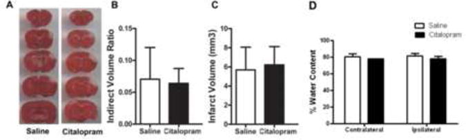
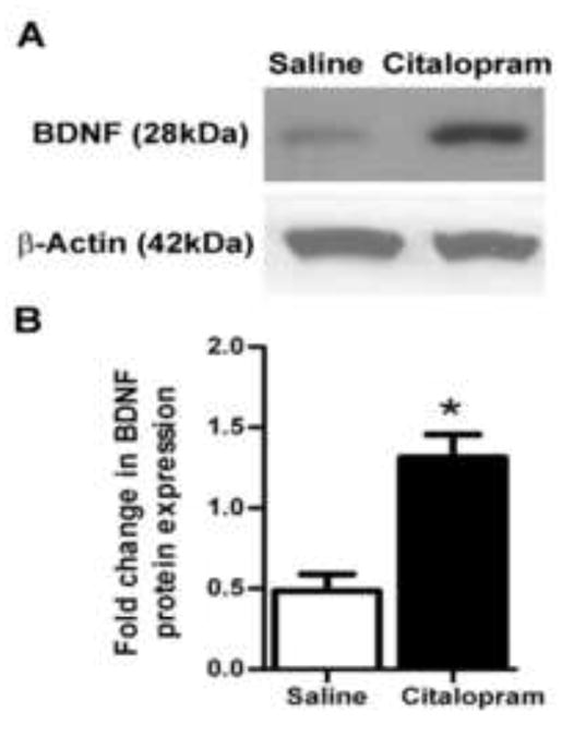
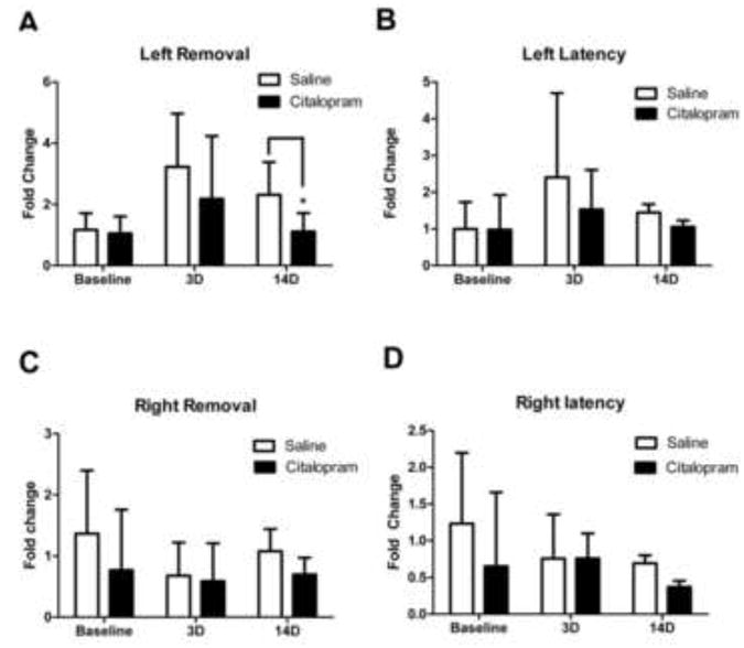
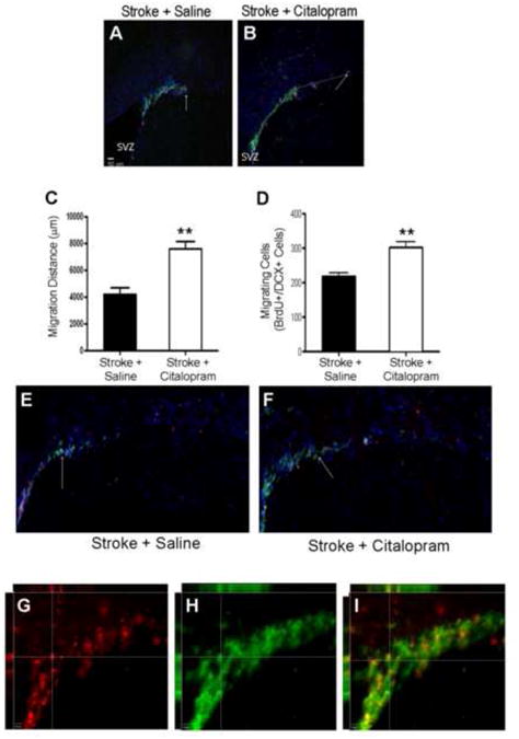
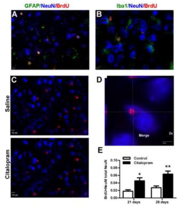
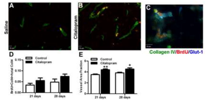
Recent clinical trials have demonstrated that treatment with selective serotonin reuptake inhibitors after stroke enhances motor functional recovery; however, the underlying mechanisms remain to be further elucidated. We hypothesized that daily administration of the clinical drug citalopram would produce these functional benefits via enhancing neurovascular repair in the ischemic peri-infarct region. To test this hypothesis, focal ischemic stroke was induced in male C57/B6 mice by permanent ligation of distal branches of the middle cerebral artery to the barrel cortex and 7-min occlusion of the bilateral common carotid arteries. Citalopram (10mg/kg, i.p.) was injected 24h after stroke and daily thereafter. To label proliferating cells, bromo-deoxyuridine was injected daily beginning 3 days after stroke. Immunohistochemical and functional assays were performed to elucidate citalopram-mediated cellular and sensorimotor changes after stroke. Citalopram treatment had no significant effect on infarct formation or edema 3 days after stroke; however, citalopram-treated mice had better functional recovery than saline-treated controls 3 and 14 days after stroke in the adhesive removal test. Increased expression of brain-derived neurotrophic factor was detected in the peri-infarct region 7 days after stroke in citalopram-treated animals. The number of proliferating neural progenitor cells and the distance of neuroblast migration from the sub-ventricular zone toward the ischemic cortex were significantly greater in citalopram-treated mice at 7 days after stroke. Immunohistochemical staining and co-localization analysis showed that citalopram-treated animals generated more new neurons and microvessels in the peri-infarct region 21 and 28 days after stroke. Taken together, these results suggest that citalopram promotes post-stroke sensorimotor recovery likely via enhancing neurogenesis, neural cell migration and the microvessel support in the peri-infarct region of the ischemic brain.
最近的临床试验表明,中风后使用选择性 5-羟色胺再摄取抑制剂治疗可以促进运动功能恢复;然而,其潜在机制仍有待进一步阐明。我们假设,临床药物西酞普兰的每日给药将通过增强缺血性梗死周围区域的神经血管修复来产生这些功能益处。为了验证这一假设,雄性 C57/B6 小鼠通过永久性结扎大脑中动脉的远端分支到大脑皮层和双侧颈总动脉 7 分钟闭塞来诱导局灶性缺血性中风。中风后 24 小时内给予西酞普兰(10mg/kg,腹腔注射),并每日给药。为了标记增殖细胞,从中风后第 3 天开始每天注射溴脱氧尿苷。进行免疫组织化学和功能测定,以阐明中风后西酞普兰介导的细胞和感觉运动变化。西酞普兰治疗对中风后 3 天的梗死形成或水肿没有显著影响;然而,在粘附去除试验中,中风后 3 天和 14 天,西酞普兰治疗组的小鼠比盐水对照组的小鼠有更好的功能恢复。中风后 7 天,西酞普兰处理的动物在梗死周围区域检测到脑源性神经营养因子的表达增加。中风后 7 天,西酞普兰治疗组的神经祖细胞增殖数量和神经母细胞从侧脑室向缺血皮层迁移的距离明显大于盐水对照组。免疫组织化学染色和共定位分析显示,西酞普兰处理的动物在中风后 21 天和 28 天在梗死周围区域产生更多的新神经元和微血管。总之,这些结果表明,西酞普兰通过促进神经发生、神经细胞迁移和缺血性脑梗死周围区域的微血管支持,促进中风后的感觉运动恢复。