Epilepsy Society MRI Unit, Chesham Lane, Chalfont St Peter, United Kingdom.
Epilepsia. 2013 Jul;54(7):1143-53. doi: 10.1111/epi.12193. Epub 2013 Apr 24.
Temporal lobe epilepsy (TLE) has been considered to impair long-term memory, whilst not affecting working memory, but recent evidence suggests that working memory is compromised. Functional MRI (fMRI) studies demonstrate that working memory involves a bilateral frontoparietal network the activation of which is disrupted in hippocampal sclerosis (HS). A specific role of the hippocampus to deactivate during working memory has been proposed with this mechanism faulty in patients with HS. Structural correlates of disrupted working memory in HS have not been explored.
We studied 54 individuals with medically refractory TLE and unilateral HS (29 left) and 28 healthy controls. Subjects underwent 3T structural MRI, a visuospatial n-back fMRI paradigm and diffusion tensor imaging (DTI). Working memory capacity assessed by three span tasks (digit span backwards, gesture span, motor sequences) was combined with performance in the visuospatial paradigm to give a global working memory measure. Gray and white matter changes were investigated using voxel-based morphometry and voxel-based analysis of DTI, respectively.
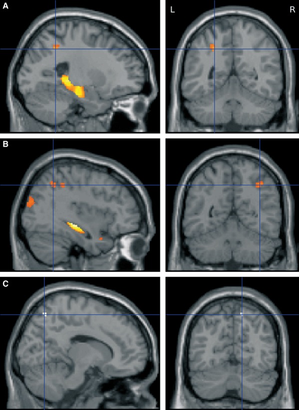
Individuals with left or right HS performed less well than healthy controls on all measures of working memory. fMRI demonstrated a bilateral frontoparietal network during the working memory task with reduced activation of the right parietal lobe in both patient groups. In left HS, gray matter loss was seen in the ipsilateral hippocampus and parietal lobe, with maintenance of the gray matter volume of the contralateral parietal lobe associated with better performance. White matter integrity within the frontoparietal network, in particular the superior longitudinal fasciculus and cingulum, and the contralateral temporal lobe, was associated with working memory performance. In right HS, gray matter loss was also seen in the ipsilateral hippocampus and parietal lobe. Working memory performance correlated with the gray matter volume of both frontal lobes and white matter integrity within the frontoparietal network and contralateral temporal lobe.
Our data provide further evidence that working memory is disrupted in HS and impaired integrity of both gray and white matter is seen in functionally relevant areas. We suggest this forms the structural basis of the impairment of working memory, indicating widespread and functionally significant structural changes in patients with apparently isolated HS.
颞叶癫痫(TLE)被认为会损害长期记忆,而不会影响工作记忆,但最近的证据表明工作记忆受到了损害。功能磁共振成像(fMRI)研究表明,工作记忆涉及一个双侧额顶叶网络,其激活在海马硬化(HS)中受到干扰。有人提出,在工作记忆期间,海马体需要去激活,而这一机制在 HS 患者中存在缺陷。HS 中工作记忆受损的结构相关性尚未得到探索。
我们研究了 54 名药物难治性 TLE 伴单侧 HS(29 例左侧)和 28 名健康对照者。受试者接受了 3T 结构磁共振成像、视觉空间 n-back fMRI 范式和弥散张量成像(DTI)检查。通过三个跨度任务(数字跨度倒转、手势跨度、运动序列)评估工作记忆能力,并将其与视觉空间范式的表现相结合,得出整体工作记忆测量值。使用基于体素的形态测量法和基于体素的 DTI 分析分别研究灰质和白质变化。
左或右 HS 患者在所有工作记忆测量中均表现不如健康对照组。fMRI 显示,在工作记忆任务中存在双侧额顶叶网络,两组患者的右侧顶叶激活减少。在左侧 HS 中,同侧海马体和顶叶出现灰质丢失,而对侧顶叶灰质体积保持不变与更好的表现相关。额顶叶网络内的白质完整性,特别是上纵束和扣带束,以及对侧颞叶与工作记忆表现相关。在右侧 HS 中,同侧海马体和顶叶也出现灰质丢失。工作记忆表现与双侧额叶的灰质体积以及额顶叶网络和对侧颞叶内的白质完整性相关。
我们的数据进一步证明,工作记忆在 HS 中受到损害,并且在功能相关区域中观察到灰质和白质的完整性受损。我们认为这构成了工作记忆受损的结构基础,表明在明显孤立的 HS 患者中存在广泛的、具有功能意义的结构变化。