Developmental and Stem Cell Biology, The Hospital for Sick Children Research Institute, Toronto, ON, Canada.
PLoS One. 2013 Apr 19;8(4):e61340. doi: 10.1371/journal.pone.0061340. Print 2013.
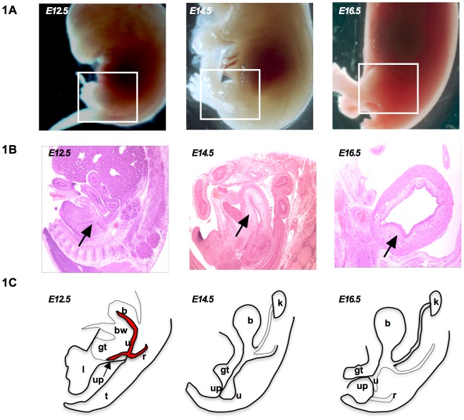
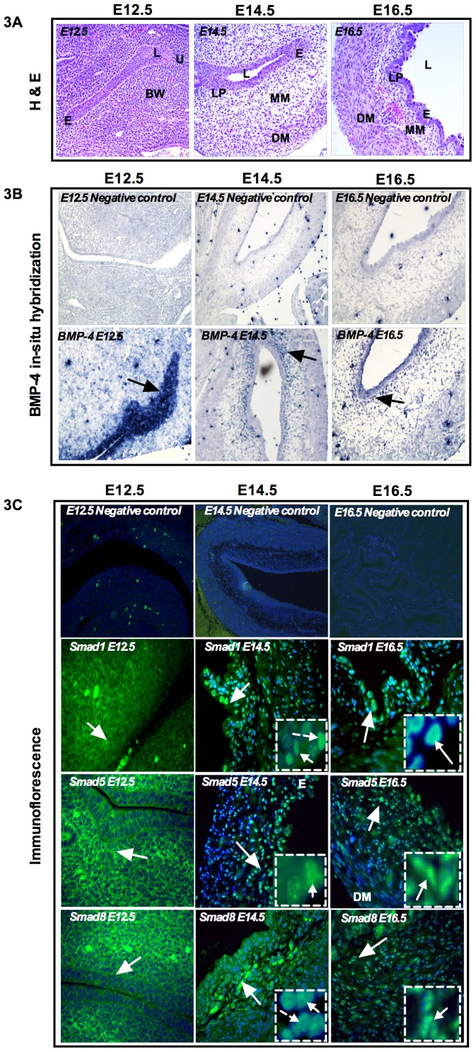
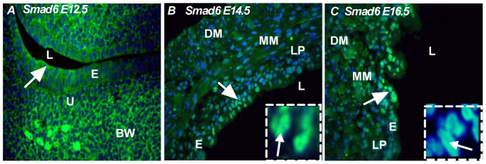
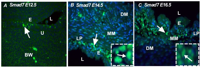
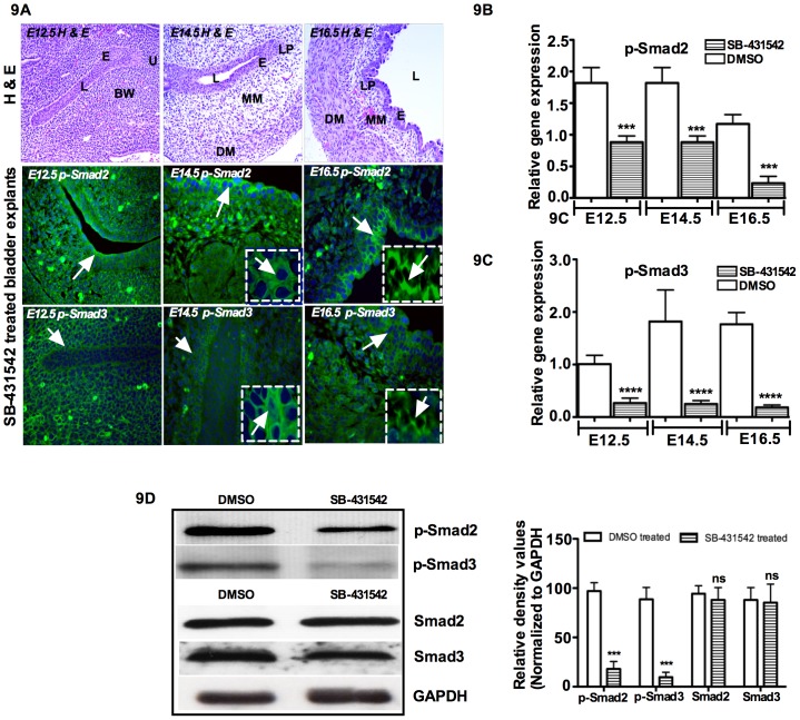
Although Shh, TGF-β and BMP-4 regulate radial patterning of the bladder mesenchyme and smooth muscle differentiation, it is not known what transcription factors, local environmental cues or signaling cascades mediate bladder smooth muscle differentiation. We investigated the expression patterns of signaling mediated by Smad2 and Smad3 in the mouse embryonic bladder from E12.5 to E16.5 by using qRT-PCR, in situ hybridization and antibodies specifically recognizing individual Smad proteins. The role of Smad2 and Smad3 during smooth muscle formation was examined by disrupting the Smad2/3 signaling pathway using TβR1 inhibitor SB-431542 in organ culture system. qRT-PCR results showed that R-Smads, Co-Smad and I-Smads were all expressed during bladder development. RNA ISH for BMP-4 and immunostaining of TGF-β1 showed that BMP-4 and TGF-β1 were expressed in the transitional epithelium, lamina propia and muscularis mucosa. Smad1, Smad5 and Smad8 were first expressed in the bladder epithelium and continued to be expressed in the transitional epithelium, muscularis mesenchyme and lamina propia as the bladder developed. Smad2, Smad3 and Smad4 were first detected in the bladder epithelium and subsequently were expressed in the muscularis mesenchyme and lamina propia. Smad6 and Smad7 showed overlapping expression with R-Smads, which are critical for bladder development. In bladder explants (E12.5 to E16.5) culture, Smad2 and Smad3 were found localized within the nuclei, suggesting critical transcriptional regulatory effects during bladder development. E12.5 to E16.5 bladders were cultured with and without TβR1 inhibitor SB-431542 and assessed by qRT-PCR and immunofluorescence. After three days in culture in SB-431542, α-SMA, Smad2 and Smad3 expressions were significantly decreased compared with controls, however, with no significant changes in the expression of smooth muscle myosin heavy chain (SM-Myh. Based on the Smad expression patterns, we suggest that individual or combinations of Smads may be necessary during mouse bladder organogenesis and may be critical mediators for bladder smooth muscle differentiation.
虽然 Shh、TGF-β 和 BMP-4 调节膀胱间质的放射状模式和平滑肌分化,但尚不清楚哪些转录因子、局部环境线索或信号级联介导膀胱平滑肌分化。我们通过 qRT-PCR、原位杂交和特异性识别单个 Smad 蛋白的抗体,研究了 Smad2 和 Smad3 介导的信号在 E12.5 至 E16.5 小鼠胚胎膀胱中的表达模式。通过在器官培养系统中使用 TβR1 抑制剂 SB-431542 破坏 Smad2/3 信号通路,研究了 Smad2 和 Smad3 在平滑肌形成中的作用。qRT-PCR 结果表明,R-Smads、Co-Smad 和 I-Smads 在膀胱发育过程中均有表达。BMP-4 的 RNA ISH 和 TGF-β1 的免疫染色显示,BMP-4 和 TGF-β1 表达于过渡上皮、固有层和黏膜肌层。Smad1、Smad5 和 Smad8 首先在膀胱上皮中表达,并随着膀胱的发育继续在过渡上皮、肌间质和固有层中表达。Smad2、Smad3 和 Smad4 首先在膀胱上皮中检测到,随后在肌间质和固有层中表达。Smad6 和 Smad7 与 R-Smads 重叠表达,对膀胱发育至关重要。在膀胱外植体(E12.5 至 E16.5)培养中,发现 Smad2 和 Smad3 定位于核内,表明在膀胱发育过程中具有关键的转录调节作用。E12.5 至 E16.5 膀胱在有和没有 TβR1 抑制剂 SB-431542 的情况下培养,并通过 qRT-PCR 和免疫荧光进行评估。在 SB-431542 培养三天后,与对照组相比,α-SMA、Smad2 和 Smad3 的表达明显降低,但平滑肌肌球蛋白重链(SM-Myh)的表达没有明显变化。基于 Smad 的表达模式,我们认为单个或组合的 Smad 可能是小鼠膀胱器官发生所必需的,并且可能是膀胱平滑肌分化的关键介质。