Department of Respiratory Medicine, Zhongshan Hospital, Fudan University, Shanghai 200032, China.
J Cell Mol Med. 2013 Apr;17(4):567-77. doi: 10.1111/jcmm.12052.
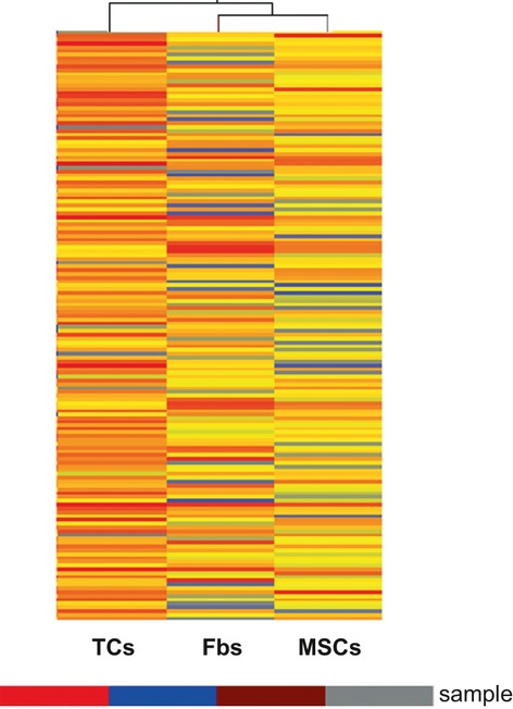
Telocytes (TCs) are interstitial cells with telopodes - very long prolongations that establish intercellular contacts with various types of cells. Telocytes have been found in many organs and various species and have been characterized ultrastructurally, immunophenotypically and electrophysiologically (www.telocytes.com). Telocytes are distributed through organ stroma forming a three-dimensional network in close contacts with blood vessels, nerve bundles and cells of the local immune system. Moreover, it has been shown that TCs express a broad range of microRNAs, such as pro-angiogenic and stromal-specific miRs. In this study, the gene expression profile of murine lung TCs is compared with other differentiated interstitial cells (fibroblasts) and with stromal stem/progenitor cells. More than 2000 and 4000 genes were found up- or down-regulated, respectively, in TCs as compared with either MSCs or fibroblasts. Several components or regulators of the vascular basement membrane are highly expressed in TCs, such as Nidogen, Collagen type IV and Tissue Inhibitor of Metalloproteinase 3 (TIMP3). Given that TCs locate in close vicinity of small vessels and capillaries, the data suggest the implication of TCs in vascular branching. Telocytes express also matrix metalloproteases Mmp3 and Mmp10, and thus could regulate extracellular matrix during vascular branching and de novo vessel formation. In conclusion, our data show that TCs are not fibroblasts, as the ultrastructure, immunocytochemistry and microRNA assay previously indicated. Gene expression profile demonstrates that TCs are functionally distinct interstitial cells with specific roles in cell signalling, tissue remodelling and angiogenesis.
间质细胞(TCs)是具有长突起的细胞,通过这些突起与各种类型的细胞建立细胞间接触。TCs 已经在许多器官和各种物种中被发现,并通过超微结构、免疫表型和电生理学进行了特征描述(www.telocytes.com)。TCs 分布在器官基质中,形成与血管、神经束和局部免疫系统细胞紧密接触的三维网络。此外,已经表明 TCs 表达广泛的 microRNAs,如促血管生成和基质特异性 miRs。在这项研究中,比较了小鼠肺 TCs 的基因表达谱与其他分化的间质细胞(成纤维细胞)和基质干细胞/祖细胞。与 MSC 或成纤维细胞相比,TCs 中分别有超过 2000 个和 4000 个基因上调或下调。TCs 中高度表达几种血管基底膜的组成部分或调节剂,如 Nidogen、IV 型胶原和基质金属蛋白酶组织抑制剂 3(TIMP3)。鉴于 TCs 位于小血管和毛细血管附近,这些数据表明 TCs 参与了血管分支。TCs 还表达基质金属蛋白酶 MMP3 和 MMP10,因此可以在血管分支和新血管形成过程中调节细胞外基质。总之,我们的数据表明 TCs 不是成纤维细胞,正如先前的超微结构、免疫细胞化学和 microRNA 检测所表明的那样。基因表达谱表明 TCs 是具有特定细胞信号转导、组织重塑和血管生成作用的功能独特的间质细胞。